Notes
Topics Covered
|
Introduction — What Is a Tissue? :
| 📘 Tissue (Definition) : A tissue is a group of cells, similar in structure, that work together to perform a specific function. |
Life begins with a single cell that divides repeatedly to form many cells. In multicellular organisms, these cells specialise and group together, giving rise to a hierarchy of organisation:
Cell → Tissue → Organ → Organ System → Organism
- Unicellular organisms (e.g., Amoeba) — a single cell performs all functions of life.
- Multicellular organisms (plants, animals) — different groups of cells (tissues) perform different functions. This is called division of labour, which increases efficiency and allows complex life processes.
Examples of Division of Labour :
- Animals: Muscle tissue enables movement; nervous tissue carries messages.
- Plants: Xylem transports water and minerals; phloem transports food.
Why Are Plant and Animal Tissues Different? :
Plant and animal tissues differ because plants and animals differ fundamentally in lifestyle, structure, and nutrition.
| Basis of Difference | Plants | Animals |
| Movement / Locomotion | Mostly fixed in one place; need support to stay upright | Mostly mobile (a few like sponges are immobile) |
| Cell wall | Present — provides rigidity and strength | Absent — allows cells to change shape easily, aiding locomotion |
| Mode of nutrition | Autotrophic — tissues help utilise solar energy via photosynthesis | Heterotrophic — tissues help digest food from external sources |
| Growth pattern | Growth continues throughout life at specific zones (meristems) | Growth occurs mainly up to a certain age, more uniformly |
| Transport tissues | Xylem (water/minerals) and phloem (food) | Blood and lymphatic tissue |
Tissues for Growth in Plants :
Plants grow in three distinct ways, each driven by a special group of actively dividing cells called meristematic tissue:
- Increase in length — height of stem and depth of roots
- Increase in girth — thickness of the stem
- Regrowth — after cutting of branches or grazing by animals
| 📘 Meristematic Tissue (Definition) : A tissue made of actively and continuously dividing cells, responsible for growth in plants. |
Characteristics of Meristematic Cells :
- Small in size, with thin cell walls
- Large, prominent nucleus and dense cytoplasm with many organelles
- Vacuoles generally absent
- Cells tightly packed with little or no intercellular space
These features allow continuous and rapid cell division.
Three Types of Meristematic Tissue :
(i) Apical Meristem :
- Apical meristem is found at the tips of roots and shoots.
- Its cells divide continuously and help the root grow deeper into the soil and the shoot grow upward.
- If the root tip is cut off, the root stops growing in length.
- Therefore, apical meristem is responsible for the growth in length of the plant.
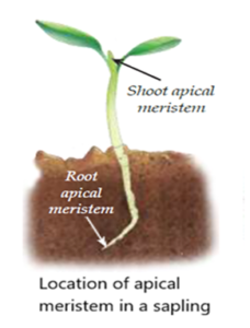
| Onion Root Experiment
• Roots grow only from their tips, which contain actively dividing (meristematic) cells — confirmed earlier by observing mitosis in onion root tips. |
(ii) Lateral Meristem :
- Lateral meristem is found along the sides or circumference of the stem.
- Its cells divide and add new cells, making the stem wider or thicker.
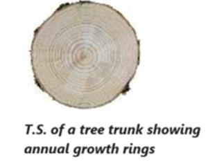
- The rings seen in a cross-section of a tree trunk are called annual growth rings.
- Each ring generally represents one year of growth.
- Wide rings show good growing conditions, while narrow rings show poor or dry conditions.
- By counting these rings, scientists can estimate the age of a tree and learn about past climate conditions.
- Therefore, lateral meristem is responsible for growth in girth or thickness.
(iii) Intercalary Meristem :
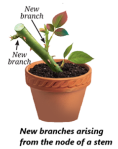
- Intercalary meristem is found at the base of an internode or just above a node.
- A node is the point on a stem where leaves or branches arise.
- The part of the stem between two nodes is called an internode.
- Intercalary meristem helps plants grow again after their tips are cut.
- For example, grass grows back quickly after mowing because of intercalary meristem.
- It also helps trimmed plants and hedges produce new branches.
- Therefore, intercalary meristem is responsible for regrowth after cutting or grazing.
Difference between Apical, Lateral and Intercalary Meristem :
| Type | Location | Function |
| Apical meristem | Tips of roots and shoots | Increases length of the plant (root grows deeper, shoot grows taller) |
| Lateral meristem | Arranged in a ring around the stem (cambium) | Increases girth/diameter of the stem; forms annual growth rings |
| Intercalary meristem | Base of internode, just above the node | Helps plant regenerate/regrow after cutting or grazing (e.g., grass after mowing) |
Differentiation :
- Meristematic tissue continuously produces new cells by cell division.
- Some of these new cells remain meristematic and continue to divide.
- Other cells stop dividing and undergo changes in their structure and shape.
- These cells become specialised to perform specific functions such as support, transport, or storage.
- This process of changing cells into specialised cells is called differentiation.
- The cells that have differentiated and can no longer divide are called permanent tissue.
| 📘 Differentiation (Definition) : The process by which meristematic tissue becomes specialised (in structure and function) to form permanent tissue. Meristematic tissue becomes permanent through differentiation. |
Permanent Tissues in Plants :
Cells that lose the ability to divide undergo differentiation and become permanent tissues, specialised for functions such as protection, support, or transport.
Permanent Tissue — Two Categories
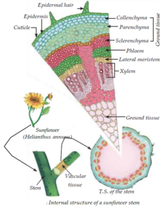
Simple permanent tissue: composed of only ONE type of cell (e.g., parenchyma, collenchyma, sclerenchyma).
Complex permanent tissue: composed of MORE THAN ONE type of cell working together (e.g., xylem, phloem).
(i) Protective Tissue — Epidermis
- Epidermis is the outermost layer of the plant body.
- It acts as the first line of defence and protects the plant.
- It is usually made up of a single layer of tightly packed cells with no spaces between them.
- The cells are covered with a waxy layer called the cuticle, which contains a substance called cutin.
Functions of Epidermis
Reduces Water Loss : The cuticle reduces excessive loss of water from the plant.
Plants growing in dry areas usually have a thicker cuticle.
Protection : The epidermis protects the plant from mechanical damage. It also helps prevent the entry of disease-causing organisms.
Root Hairs : In roots, epidermal cells form long, hair-like structures called root hairs. Root hairs increase the surface area of the root and help absorb water and minerals from the soil.
Stomata :
- Leaves have tiny pores called stomata in the epidermis.
- Each stoma is surrounded by two guard cells, which control its opening and closing.
- Stomata allow the exchange of gases needed for photosynthesis and respiration.
- They also help in transpiration, which is the loss of water vapour from leaves.
- Transpiration creates a transpiration pull that helps draw water upward through the xylem.
| 💡 Know This : How is Bark Formed?
· In young plants, the outer protective layer is a single-layered epidermis. · As the plant grows older, cells just below the epidermis develop the ability to divide again, forming a new layer called the cork cambium. · The cork cambium produces cork cells on the outside. These cells are dead, tightly packed, and impermeable to water and gases. · This protective layer of cork cells forms the bark seen on the outside of old tree trunks. |
(ii) Simple Permanent Tissues :
- Simple permanent tissues are made up of similar types of cells.
- They provide support, strength, storage, and flexibility to the plant.
There are three main types: parenchyma, collenchyma, and sclerenchyma.
(a) Parenchyma :
- Parenchyma is the most common type of simple permanent tissue.
- It is found in the soft parts of plants such as leaves, roots, fruits, and flowers.
- It consists of living cells with thin cell walls.
- The cells are usually loosely packed, leaving spaces between them.
- Parenchyma fills spaces between other tissues and helps maintain the shape of the plant.
- It also stores food and water.
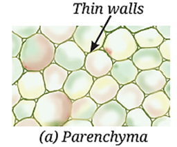
Types of Parenchyma
- Chlorenchyma : It contains chlorophyll. It helps the plant perform photosynthesis.
- Aerenchyma : It contains large air spaces. It provides buoyancy to aquatic plants and helps them float.
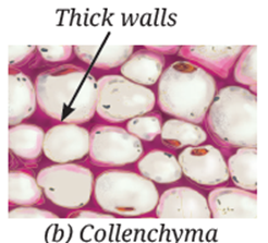
(b) Collenchyma :
- Collenchyma consists of living cells with unevenly thickened corners.
- The thickening is mainly due to the deposition of pectin.
- It provides strength and flexibility to young plant parts.
- It allows the plant parts to bend without breaking.
- It is mainly found in young stems and leaf stalks.
- For example, the stalk of a coriander leaf is flexible because of collenchyma.
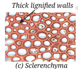
(c) Sclerenchyma :
- Sclerenchyma consists of cells with very thick and hard walls.
- The walls contain a substance called lignin, which makes them strong and rigid.
- Most sclerenchyma cells are dead at maturity.
- They provide hard and strong support to the plant.
- They are found in stems, leaf veins, and hard seed coats.
- Examples include the coconut husk and walnut shell.
Differences between Parenchyma, Collenchyma and Sclerenchyma :
| Tissue | Cell Nature | Wall / Structure | Function | Example Location |
| Parenchyma | Living | Thin walls; loosely packed with intercellular spaces | Stores food; photosynthesis in green parts; forms air spaces for floating in aquatic plants | Soft parts — pith, leaves, fruit pulp |
| Collenchyma | Living | Unevenly thickened corners (pectin deposition) | Provides flexibility & support — lets stems/tendrils bend without breaking | Stems, leaf stalks (e.g., coriander) |
| Sclerenchyma | Mostly dead | Thick walls due to lignin deposition — hard & strong | Provides mechanical strength/rigidity | Stems, leaf veins, seed & nut coats (coconut husk, walnut shell) |
(iii) Complex Permanent (Conducting) Tissues :
- Complex permanent tissues are made up of different types of cells that work together to perform a common function.
- Plants have two main types of complex permanent tissues: xylem and phloem.
- Together, xylem and phloem are called vascular tissues.
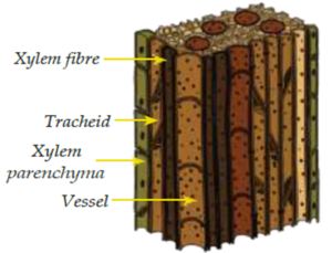
(a) Xylem : Xylem transports water and dissolved minerals from the roots to all parts of the plant. It also provides mechanical strength and support to the plant.
Xylem consists of four types of cells.
- (i) Tracheids : Tracheids are long, narrow and dead cells with thick walls. They help transport water through small openings in their walls.
- (ii) Vessels : Vessels are wide, dead cells joined end to end. They form long tubes through which water can flow. They transport water faster and more efficiently than tracheids.
- (iii) Xylem Parenchyma : Xylem parenchyma is the only living tissue in xylem. It stores food. It also helps in the sideways movement of water.
- (iv) Xylem Fibres : Xylem fibres are dead cells with thick walls. They provide strength and support to the plant.
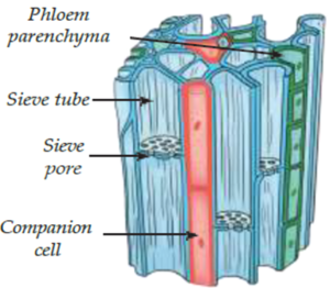
(b) Phloem : Phloem transports food, mainly sugars made in the leaves, to different parts of the plant. It transports food to parts such as the roots, stems, fruits, and growing regions. Unlike xylem, phloem is mainly made up of living cells.
Phloem consists of four types of cells.
- (i) Sieve Tubes : Sieve tubes are long cells joined end to end. Their walls have small openings that look like a sieve. Food moves through these tubes from the leaves to other parts of the plant.
- (ii) Companion Cells : Companion cells are specialised living cells closely associated with sieve tubes. They help in the loading and unloading of sugars into and out of the sieve tubes.
- (iii) Phloem Parenchyma : Phloem parenchyma consists of living cells. They store food and substances such as resin, tannins, and latex.
- (iv) Phloem Fibres : Phloem fibres are dead cells with thick walls. They provide strength and support to the phloem.
| 💡 Know This : Totipotency :
The ability of a single mature cell to develop into a complete organism is called totipotency. Cells that have this ability are called totipotent cells.
|
Differences between Xylem and Phloem :
| Feature | Xylem | Phloem |
| Function | Transports water & minerals from roots to rest of plant; also provides mechanical strength | Transports food (prepared in leaves) to all parts of the plant |
| Direction of transport | Upward (roots → shoot/leaves) | Both directions (source → sink) |
| Made up of | Tracheids, vessels, xylem parenchyma, xylem fibres | Sieve tubes, companion cells, phloem parenchyma, phloem fibres |
| Living / Dead | Mostly dead (tracheids, vessels, fibres); only xylem parenchyma is living | Mostly living (except phloem fibres, which are sclerenchymatous) |
| Special cells | Tracheids & vessels — tubular, thick-walled, for water conduction | Sieve tubes — long tubular cells joined end-to-end by perforated (sieve) walls; regulated by companion cells |
(iv) Tissue Systems in Plants :
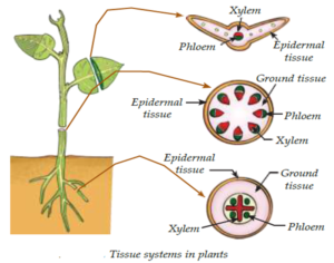
In a plant body, tissues are not scattered randomly. They are organised into three larger groups called tissue systems, each covering the entire plant from roots to leaves.
| Tissue System | Composition | Function |
| Dermal tissue system | Epidermis (+ cuticle) | Forms outer covering; protects inner parts and reduces water loss |
| Ground tissue system | Parenchyma, collenchyma, sclerenchyma | Forms the main body of the plant between dermal and conducting tissues |
| Vascular tissue system | Xylem and phloem | Conducts water, minerals, and food throughout the plant |
Animal Tissues :
Like plants, animal cells group together and specialise to form tissues. Animal tissues help in important life processes such as movement, digestion, communication and protection.
There are four main types of animal tissue:
- Epithelial tissue — covering/lining and protection
- Connective tissue — connects and supports
- Muscular tissue — movement
- Nervous tissue — control and coordination
| 🧠 Remember the 4 Animal Tissues — "E-C-M-N"
"Every Cat Moves Nimbly" → Epithelial, Connective, Muscular, Nervous. |
(1) Epithelial Tissue
| 📘 Epithelial Tissue (Definition) : Forms the outer covering of the body (skin) and lines internal organs (mouth, lungs, blood vessels, intestine).
· Cells are closely packed with very little intercellular space, which prevents germ entry, reduces water loss, and allows absorption, secretion, and movement of substances. |
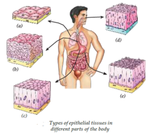
Types of Epithelial Tissue :
Different types of epithelial tissues have different structures and functions.
(a) Gaseous and Liquid Diffusion :
- This tissue has a single layer of thin, flat cells. These cells allow substances to pass through them easily.
- It is found in the lining of blood vessels and lungs, where the exchange of gases and other substances takes place.
(b) Protective Lining :
- This tissue has many layers of tightly packed, flat cells. These layers provide strength and protect the body from injury, friction and germs.
- It is found in the skin, mouth and oesophagus.
(c) Secretory Tissue :
- This tissue helps in the secretion of substances such as mucus, enzymes, hormones and saliva.
- Its cells are usually cuboidal or columnar in shape. It is found in the salivary glands, sweat glands and stomach lining.
(d) Sensory Epithelium :
- This tissue helps the body detect sensory stimuli such as smell, taste, sound and balance.
- It contains special receptor cells, often with hair-like projections. It is found in the nostrils, taste buds and inner ear.
(e) Absorptive Lining :
- This tissue consists of a single layer of tall, pillar-shaped cells. These cells have tiny projections called microvilli, which increase the surface area for absorption.
- It helps in the absorption of nutrients and water and is mainly found in the lining of the small intestine.
(2) Connective Tissues :
Connective tissue connects, supports and holds together different tissues and organs of the body. Its cells are present in a non-living material called the matrix. The nature of the matrix gives each connective tissue its special properties.
Main Types of Connective Tissue
The main types are:
- Blood
- Bone
- Cartilage
- Tendons
- Ligaments
(i) Blood : Blood is a fluid connective tissue. Its liquid matrix is called plasma. Plasma makes up about 55% of blood. It transports nutrients, hormones, gases and waste products throughout the body.
- Red Blood Cells (RBCs) : RBCs contain haemoglobin, an iron-rich protein that gives blood its red colour. They carry oxygen to all parts of the body. They live for about four months.
- White Blood Cells (WBCs) : WBCs help protect the body from infections and diseases. They are an important part of the body's immune system. They gather at infected areas and help fight germs.
- Platelets : Platelets are small cell fragments present in blood. They help in blood clotting when an injury occurs. They prevent excessive loss of blood.
(ii) Bone : Bone is a connective tissue with a matrix rich in calcium and phosphorus.
- This mineral-rich matrix makes bones strong and hard.
- Bones provide support and shape to the body.
- They protect important organs such as the brain, heart and lungs.
(iii) Cartilage : Cartilage has a soft and flexible matrix.
- It provides flexibility and cushioning.
- It is found at the ends of bones, in the joints, ear, nose and between the vertebrae.
- It acts as a shock absorber and protects the ends of bones during movement.
(iv) Tendons : Tendons are strong and flexible bands of connective tissue.
- They connect muscles to bones.
- When muscles contract, tendons transmit the force to bones.
- This helps produce movement at joints.
- Tendons can withstand strong pulling forces.
(v) Ligaments : Ligaments are strong and slightly elastic connective tissues.
- They connect one bone to another bone at joints.
- They provide stability to joints.
- They prevent excessive movement and help protect joints from dislocation.
- Their slight elasticity allows controlled movement while keeping the joint stable.
| 💡 Quick Distinguish — Tendon vs Ligament
• Tendon: Muscle → Bone ("Tendon = Toward bone from muscle") • Ligament: Bone → Bone ("Ligament = Links bones together") |
Quick Overview :
| Type | Matrix | Function | Example / Location |
| Blood | Fluid (plasma) | Transports nutrients, gases, hormones, wastes throughout the body | Circulatory system (RBCs, WBCs, platelets, plasma) |
| Bone | Hard, solid, rigid (calcium & phosphorus compounds) | Gives strength, support, and protection | Skeleton — collar bone, long bones, kneecap |
| Cartilage | Soft, jelly-like — flexible | Provides flexibility; cushions bone ends for shock absorption | Ear, nose, joints, between vertebrae |
| Tendon | Tough fibrous connective tissue | Connects muscle to bone; brings about movement | Around joints, e.g., ankle (Achilles tendon) |
| Ligament | Tough, slightly elastic connective tissue | Connects bone to bone; provides stability, limits movement, prevents dislocation | Around joints, e.g., knee |
(3) Muscular Tissue :
Muscular tissue is made up of special cells called muscle fibres. It helps the body and its organs to move. There are three main types of muscular tissue:
- Skeletal muscle
- Smooth muscle
- Cardiac muscle
(i) Skeletal Muscle (Voluntary Muscle)
- Skeletal muscle is also called striated muscle because it has light and dark bands called striations.
- It is made up of long, cylindrical and unbranched fibres.
- Each fibre has many nuclei.
- These muscles are attached to bones through tendons.
- Their movement is under our conscious control.
- They help us perform activities such as walking, running, writing and lifting.
- They may fatigue quickly after prolonged use.
(ii) Smooth Muscle (Involuntary Muscle)
- Smooth muscle is also called unstriated muscle because it does not have striations.
- Its cells are spindle-shaped and have a single nucleus.
- These muscles are found in internal organs such as the stomach, intestines and blood vessels.
- Their movement is not under our conscious control.
- They help in activities such as moving food through the intestine and controlling blood flow.
- They produce slow, continuous and sustained contractions.
(iii) Cardiac Muscle :
- Cardiac muscle is found only in the walls of the heart.
- Its cells are cylindrical and branched.
- They usually have a single nucleus and faint striations.
- Their contractions are involuntary, rhythmic and continuous.
- They work continuously throughout life to pump blood around the body.
- Cardiac muscles are highly resistant to fatigue because they have a rich blood supply and many mitochondria.
| Type | Cell Shape / Nucleus | Striations | Control | Location |
| Skeletal (voluntary) | Long, cylindrical, unbranched, multinucleate | Striated (light & dark bands) | Voluntary | Attached to skeleton (arms, legs) |
| Smooth (involuntary) | Spindle-shaped, single nucleus | Non-striated | Involuntary | Stomach, intestines |
| Cardiac (involuntary) | Cylindrical, branched, single nucleus | Faintly striated | Involuntary | Heart only |
(4) Nervous Tissue :
Nervous tissue forms the control and coordination system of the body. It helps the body receive, process and transmit messages quickly.
- The brain acts as the control centre of the body.
- It controls activities, memory and responses.
- The spinal cord carries messages between the brain and different parts of the body.
- Muscles, whether voluntary or involuntary, receive instructions from nervous tissue.
- For example, during exercise, the brain signals the heart to beat faster to supply more oxygen to the body.
- The basic structural and functional unit of nervous tissue is called a neuron or nerve cell.
Structure of a Neuron :
A neuron has four main parts:
- Cell Body : The cell body contains the nucleus. It controls the activities of the neuron. It is the main metabolic centre of the neuron.
- Dendrites : Dendrites are short, branched extensions of the cell body. They receive signals from other neurons or sensory receptors. They carry these signals towards the cell body.
- Axon : The axon is a long fibre extending from the cell body. It carries nerve impulses away from the cell body. It passes messages to other neurons, muscles or glands.
- Axon Terminals : Axon terminals are the branched endings of the axon. They transmit signals to the next neuron or an effector organ. An effector organ may be a muscle or gland.
The Musculoskeletal System :
| 📘 Musculoskeletal System (Definition) : Made up of bones, muscles, joints, cartilage, tendons, and ligaments. |
- It is controlled by the nervous system.
- It gives support and shape to the body.
- It helps maintain body posture.
- It allows the body to move.
- It protects important internal organs.
- Muscles are attached to bones by tendons.
- When muscles contract, they pull the bones and produce movement at the joints.
- The human skeleton makes up about 12–15% of body weight.
- Muscle mass is about 40–50% in males and 30–40% in females.
Types of Movement in the Musculoskeletal System
Different parts of the body can move in different ways depending on the type of joint present.
What is a Joint?
- A joint is the place where two or more bones meet.
- Joints allow bones to move.
- A joint itself does not move the bones.
- Movement is produced when muscles contract and pull the bones.
Types of Joints and Their Movements :
Different joints allow different types of movement.
(i) Ball and Socket Joint :
- The shoulder joint allows the arm to move freely.
- The rounded end of the upper arm bone fits into a hollow in the shoulder bone.
- This forms a ball and socket joint.
- It allows the arm to move forward, backward, sideways and in a circular direction.
- The shoulder, along with the collarbone, forms the shoulder girdle, which connects the arm to the skeleton.
(ii) Hinge Joint :
- The elbow joint allows the arm to bend and straighten in one direction.
- It works like a door hinge.
- This type of joint is called a hinge joint.
- A similar joint is present in the knee.
- The knee has a small bone called the kneecap, which helps protect the joint.
(iii) Pivot Joint :
- The pivot joint allows the head to turn from side to side.
- The skull is connected to the backbone through a pivot joint in the neck.
- It allows the head to move from side to side.
- This movement is similar to a doorknob turning in its socket.
(iv) Fixed Joints :
- The bones of the skull are joined by fixed joints.
- These joints do not allow the bones to move.
- The skull forms a hard protective case around the brain, eyes and ears.
- Fixed joints help keep the brain safe and protect it from injury.
The different types of joints, their movements, locations and examples are summarised in the table below
| Joint Type | Structure | Movement Allowed | Example |
| Ball and socket | Rounded top of one bone fits into a hollow of another | Free movement — forward, backward, sideways, circular | Shoulder joint |
| Hinge | Bends and straightens in one direction only (like a door hinge) | Movement in a single plane | Elbow, knee |
| Pivot | One bone rotates within a ring formed by another (like a doorknob) | Side-to-side / rotational movement | Joint between skull and backbone (neck) |
| Fixed (immovable) | Bones joined together rigidly, cannot move | No movement | Skull bones (protect the brain) |
In Short
- Ball and socket joint → Allows movement in many directions.
- Wrist joint (Partial rotation joint): Allows circular and side to side movements of the hand.
- Hinge joint → Allows bending and straightening.
- Pivot joint → Allows rotation.
- Fixed joint → Does not allow movement.
| 🧠 Remember Joint Types — "BHPF" : "Big Horses Prefer Fences" → Ball-and-socket, Hinge, Pivot, Fixed — from most to least movement allowed. |
Skeletal System :
The skeletal system is the framework of bones that gives the body strength, shape and support. It also protects important organs.
The main parts of the skeletal system are:
- Skull
- Vertebral column
- Rib cage
(i) The Skull :
- The skull is a hard protective case made up of flat bones.
- The bones are joined by fixed joints.
- These joints do not allow movement between the skull bones.
- The skull protects important organs such as the brain, eyes and ears.
(ii) Vertebral Column (Backbone or Spine) :
- The vertebral column is a flexible column made up of small bones called vertebrae.
- It extends from the base of the skull.
- It supports the body and helps maintain an upright posture.
- It protects the spinal cord.
- Soft cartilage discs are present between the vertebrae.
- These discs act as cushions, absorb shocks and allow the body to bend and twist.
(iii) Rib Cage :
- Humans have 12 pairs of ribs that form the rib cage.
- The rib cage protects important organs such as the heart and lungs.
- Ribs are attached to the backbone at the back.
- At the front, they are connected to the sternum (breastbone) with the help of cartilage.
- This flexible arrangement allows the rib cage to expand and contract during breathing.
- It helps in inhalation and exhalation.
Yoga and Musculoskeletal Health :
- Yoga is an ancient Indian practice that includes physical postures called asanas, breathing exercises called pranayama and meditation.
- Regular yoga can improve flexibility and posture.
- It can help improve breathing and reduce stress.
- It can also help maintain healthy bones, muscles and joints.
- 21st June is observed every year as International Yoga Day.
- Correct posture, proper nutrition, regular exercise and yoga help keep bones strong, muscles fit and joints flexible throughout life.
| 💡 Exam Tip — Structure–Function Link
Whenever asked 'why does a tissue have this structure?', connect it to its function: • thin flat cells → fast diffusion; • thick lignified walls → strength; • unbranched striated fibres → strong voluntary contraction; • branched fibres with faint striations → continuous rhythmic contraction (heart). • For joints: more freedom of movement = more complex joint surface (ball-and-socket > pivot > hinge > fixed). |
| 💡 Know the Scientists:
Scientists who have made significant contributions to the fields of botany, plant anatomy, and tissue culture. B. G. L. Swamy
Sipra Guha Mukherjee and S. C. Maheshwari
F. C. Steward
|
We reply to valid query.