Notes
|
Topics to be Learn :
|
Introduction :
Nutrition and respiration are two vital biological activities that provide us energy.
Carbohydrates proteins fats vitamins minerals, water and fibres in adequate amount are the dietary needs of human being.
Nutrition : Nutrition is the sum of an organism's processes for consuming and utilising food substances.
- WHO defines nutrition as the intake of food, considered in relation to the body’s dietary needs.
Various steps involved in nutrition :
- Ingestion: It is the introduction of food into mouth, i.e. intake of food (eating) inside the body.
- Digestion: The process during which the complex, non-diffusible and non-absorbable food substances are converted into simple, diffusible and absorbable substances by the action of enzymes is called digestion.
- Absorption: The process of diffusion of digested food into blood and lymph is called absorption.
- Assimilation: The process by which protoplasm is synthesized into each cell of the body by utilizing simple food substances are called assimilation.
- Egestion: The elimination of undigested food from the body is called egestion.
Importance of Digestion :
- Digestion is a critical process that transforms complicated, non-diffusible, and non-absorbable dietary items into simple, diffusible, and assimilable compounds.
- Our food contains all essential nutrients in the form of complex components such as carbs, proteins, lipids, and vitamins.
- Through digestion, these complicated compounds are transformed into simple, diffusible, and assimilable substances.
Human Digestive System :
Digestive system of man consists of alimentary canal and associated digestive glands.
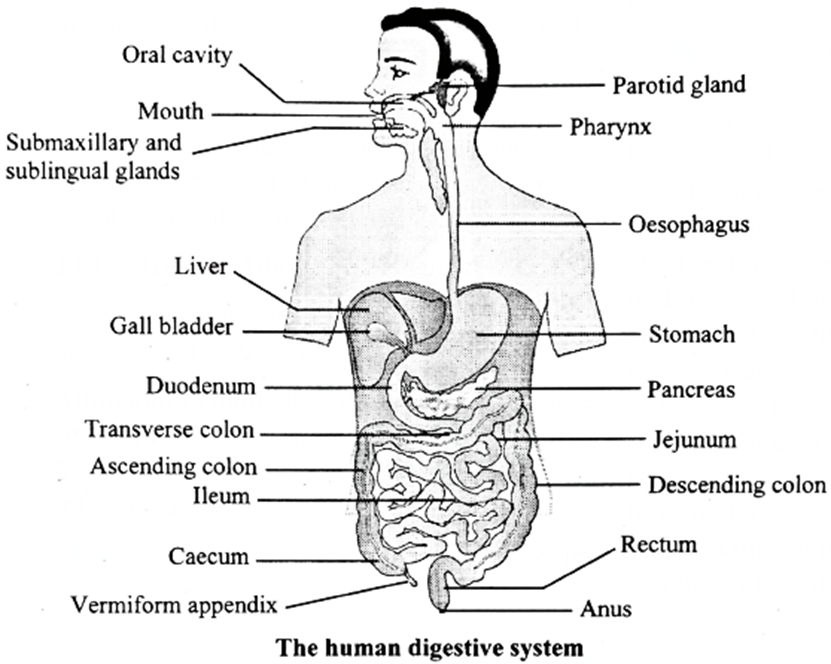
Alimentary canal :
- It is a long tubular structure starting from mouth and ending with anus. It is about 8-10 meters long and consists of following organs :
- Alimentary canal consists of mouth, buccal cavity, pharynx, oesophagus, stomach, small intestine, large intestine and anus.
(i) Mouth:
- It is also called oral or buccal cavity and is bounded by fleshy lips.
- Its side walls are formed of cheeks, roof is formed by palate and floor by tongue.
- It is internally lined by a mucous membrane.
- Salivary glands open into the buccal cavity.
- Function: It helps in ingestion of food.
(ii) Teeth
- 32 teeth are present in the buccal cavity of an adult human being.
- Human dentition is described as thecodont, diphyodont and heterodont.
- It is called thecodont type because each tooth is fixed in a separate socket present in jaw bones by gomphosis type of joint.
- In our life time, we get only two sets of teeth, milk teeth and permanent teeth. This is called diphyodont dentition.
- We have four different type of teeth hence we are heterodont.
- Types of teeth are incisors (I) canines (C) premolars (PM) and molar (M).
- Each half of each jaw has two incisors, one canine, two premolars and three molars.
Thus, dental formula of adult human can be represented as:
\(I\frac{2}{2}, C\frac{1}{1}, PM\frac{2}{2}, M\frac{3}{3} = \frac{2+1+2+3}{2+1+2+3}\) i.e. 8 x 2= 32 teeths
Structure of tooth:
- A tooth consists of the portion that projects above the gum called crown and the root that is made up of two or three projections which are embedded in gum.
- A short neck connects the crown with the root.
- The crown is covered by the hardest substance of the body called enamel which is made up of calcium phosphate and calcium carbonate.
- Basic shape of tooth is derived from dentin which is a calcified connective tissue.
- The dentin encloses the pulp cavity. It is filled with connective tissue pulp. It contains blood vessels and nerves.

- Pulp cavity has extension in the root of the tooth called root canal.
- The dentin of the root of tooth is covered by cementum which is a bone like substance that attaches the root to the surrounding socket in the gum.
Function: Teeth masticate the food into small particles and help to mix food with saliva.
Human dentition :
- Human dentition is described as thecodont, diphyodont and heterodont. .
- It is called thecodont type because each tooth is fixed in a separate socket present in the jaw bones by gomphosis type 0f joint.
- It is called diphyodont type because we get only tow sets of teeth, milk teeth and permanent teeth.
- It is called heterodont type because humans have four different type of teeth like incisors, canines, premolars and molars.
Dentition : The study of teeth with respect to their number, arrangement, development etc. is known as dentition.
(iii) Tongue:
- It is the muscular fleshy organ and is roughly triangular in shape. It lies along the floor of the buccal cavity.
- Functions: The upper surface of the tongue bears numerous projections called papillae. These papillae contain sensory receptors called taste buds. Taste buds are used to detect different tastes of the food.
- The tongue mixes saliva with food to form bolus. It helps in swallowing and in speech.
(iv) Pharynx:
- The buccal cavity leads to a short pharynx.
- Pharynx is a common passage for food and air.
- The pharynx opens into trachea through an opening called glottis.
- The glottis is guarded by a cartilaginous flap called epiglottis. The epiglottis closes during the swallowing (deglutition) action and prevents entry of food into the trachea.
- The lower region of pharynx is called oropharynx.
- Oropharynx opens into oesophagus through gullet.
- Function : Pharynx passes the food towards the oesophagus.
Know This :
|
(v) Oesophagus:
- The oesophagus is a thin, muscular tube.
- It lies behind the trachea.
- It is approximately 25cm long tube passes through the neck, central aspect of rib cage, pierces the diaphragm and joins the stomach.
- It is lined by mucus cells.
- Mucus lubricates the passageway of food.
- Oesophagus is made up of longitudinal and circular muscles.
- Function : The rhythmic wave of contraction and relaxation of these muscles is called peristalsis that helps in passage of food through oesophagus.
(vi) Stomach :
- The stomach is located in the upper left portion of the abdominal cavity.
- It is a muscular sac-like 'J' shaped organ, around 25 to 30cm in length.
- It is divided into upper cardiac region and lower pyloric region.
- Cardia or Cardiac: It is first part in which oesophagus opens. The cardia surrounds the band of circular muscles present at the junction of oesophagus and stomach called cardiac sphincter. The cardiac sphincter prevents back flow or regurgitation of food from stomach to oesophagus.

- Fundus: It is the dome shaped region above and lefi of cardia.
- Body: It forms the large central portion of stomach that stores the food.
- Pylorus: It is a narrow posterior region of stomach. It opens into duodenum, the initial pan of small intestine. This opening is guarded by a set of sphincter muscles called pyloric sphincter. It regulates the flow of food from stomach to small intestine.
- Function: The stomach temporarily stores the food. It churns the food and helps in mixing the food with gastric juice.
(vi) Small Intestine:
- It is about 6 meters long and 2.5 cm broad tube coiled within abdominal cavity.
- The coils are held together by mesenteries, supporting the blood vessels, lymph vessels and nerves.
- It is divided into three parts: Duodenum, jejunum and ileum.
- Duodenum: It is about 26 cm long ‘U’ shaped structure. The duodenum turns towards left side of abdominal cavity below the stomach
- Jejunum: It is about 2.5 meters long, coiled middle portion of small intestine. It is narrower than the duodenum.
- Ileum: It is about 3.5 meters long. It is highly coiled and little broader than jejunum. The ileum opens into the caecum of large intestine at ileocaecal junction.
- Function : In small intestine, digestion of food and absorption of nutrients take place.
(vii) Large Intestine:
- It is 1.5 meters in length. It is wider in diameter and shorter than small intestine. It consists of caecum, colon and rectum.
- Caecum: Caecum is a small, blind sac present at the junction of ileum and colon. It is 6 cm in length. It hosts some symbiotic microorganisms. An elongated worm like venniform appendix arises from the caecum. Appendix is vestigial organ in human beings and functional in herbivorous animals for the digestion of cellulose.
- Colon : Caecum opens into colon. Colon is tube like-organ consist of three parts, ascending colon, transverse colon and descending colon. The colon is internally lined by mucosal cells.
- Rectum : It is posterior region of large intestine. It temporarily stores the undigested waste material called faeces till it is egested out through anus.
(viii) Anus:
- Anus is the terminal opening of alimentary canal.
- It is guarded by sphincter.
- Function: It expels fecal matter by a process called egestion or defecation.
Differentiate between Small Intestine and Large Intestine :
| Small Intestine | Large Intestine |
| It is about 6 meters long. | It is about 1.5 meters long. |
| Small intestine is 2.5 cm broad tube. | Large intestine is broader than the small intestine. |
| It is divided into three parts, as Duodenum, Jejunum, Ileum. | It is divided into three parts as caecum, colon and rectum. |
| Absorbs the digested nutrients. | Takes part in absorption of water and minerals. |
| Vili present. | Vili absent. |
| Digestion is completed in small intestine. | No role in digestion. |
Histological Structure of Alimentary Canal :
Layers of gastrointestinal tract :
- The entire gastrointestinal tract is lined by four basic layers from inside to outside namely, mucosa, submucosa, muscularis and serosa.
- These layers show modification depending on the location and function of the organ concerned.

Serosa:
- It is the outermost layer.
- It is made up of a layer of squamous epithelium called mesothelium and inner layer of connective tissue.
Muscularis:
- This layer is formed of smooth muscles.
- These muscles are usually arranged in three concentric layers.
- Outermost layer shows longitudinal muscles, middle circular muscles and inner oblique muscles.
- This layer is wider in stomach and comparatively thin in intestinal region.
- The layer of oblique muscles is absent in the intestine.
Submucosa:
- It is formed of loose connective tissue containing blood vessels, lymph vessels and nerves.
- Duodenal submucosa shows presence of glands.
Mucosa:
- The lumen of the alimentary canal is lined by mucosa.
- - Throughout the length of alimentary canal, the mucosa layer shows presence of goblet cells that secrete mucus.
- This lubricates the lumen of alimentary canal. '
- This layer shows modification in different regions of alimentary canal. In stomach, it is thrown into irregular folds called rugae.
- In stomach mucosa layer forms gastric glands that secrete gastric juice.
Villi :
- Mucosa of small intestine forms finger like foldings called villi.
- The intestinal villi are lined by brush border or epithelial cells having microvilli at the free surface.
- Villi are supplied with a network of capillaries and lymph vessels called lacteals.
- Mucosa forms crypts in between the bases of villi in intestine called crypts of Lieberkuhn. These are intestinal glands.
Significance of the glandular epithelium of mucosa :
Globlet cells of the epithelial layer of a mucous membrane secrete mucus which lubricates the lumen 0f the elementary canal. This helps in movement of food through the gastrointestinal tract.
Digestive Glands :
The digestive glands associated with the alimentary canal include the salivary glands, liver and pancreas.
Salivary Glands:
- There are three pairs of salivary glands which open in buccal cavity.
- Parotid glands are present in front of the ear.
- The submandibular glands are present below the lower jaw.
- The glands present below the tongue are called sublingual.
- Salivary glands are made up of two types of cells.
- Serous cells secrete a fluid containing digestive enzyme called salivary amylase.
- Mucous cells produce mucus that lubricates food and helps swallowing.
Liver :
- Liver is dark reddish-brown coloured largest gland of the body, weighing 1.2 to 1.5 kg, in adults.
- Situated in right upper portion of the abdominal cavity, below the diaphragm.
- Divided into 2 lobes, right and left.
- A thin connective tissue sheath called Glisson’s capsule covers the liver and invaginates inside to divide the liver into cord like structures called hepatic lobules which are functional units of liver containing hepatic cells (hepatocytes).
- Each hepatic lobule is polygonal in shape. At the junction of adjacent lobules, a triangular portal area is present.
- In this portal area a branch of each of hepatic artery, hepatic portal vein and bile duct are present.
- Lobule consist of cords of hepatic cells which are arranged around a central vein.
- In between the cords of hepatic cells, spaces called sinusoids are present through which the blood flows. In the sinusoids, phagocytic cells called Kupffer cells are present.
- Hepatic cells secrete bile. Bile is carried by hepatic ducts in a thin muscular sac called gall bladder.
- The duct of the gall bladder and hepatic duct together form common bile duct.
- Liver synthesizes vitamins A, D, K and B12, blood proteins.

- Bile duct: It carries bile from the gall bladder and empties it into the intestine.
- Common hepatic duct: It drains bile from the liver. It helps in transportation of waste from liver and helps in digestion by releasing bile.
Pancreas:
- Pancreas is a leaf shaped heterocrine gland present in the gap formed by bend of duodenum under the stomach.
- Exocrine part of pancreas is made up of acini, the acinar cells secrete alkaline pancreatic juice that contains various digestive enzymes.
- Pancreatic juice is collected and carried to duodenum by pancreatic duct.
- The common bile duct joins pancreatic duct to fonn hepato-pancreatic duct. It opens into duodenum.
- Opening of hepato-pancreatic duct is guarded by sphincter of Oddi.
- Endocrine part of pancreas is made up of islets of Langerhans situated between the acini.
- It contains three types of cells alpha-cells which secrete glucagon, Beta-cells which secretes insulin and delta cells secrete somatostatin hormone.
- Glucagon and insulin together control the blood-sugar level.
- Somatostatin hormone inhibits glucagon and insulin secretion.

| Know This :
Importance of liver in our body.
Effects of alcohol on liver. Alcoholism causes different disorders of liver like steatosis (fatty liver), alcoholic hepatitis, fibrosis and cirrhosis. Most of the alcohol in the body is broken down in the liver by an enzyme called alcohol dehydrogenase, which transforms ethanol into a toxic compound called acetaldehyde (CH3CHO). Over consumption of alcohol leads to cirrhosis (distorted or scarred liver) and eventually to liver failure. Therefore, alcoholic people may suffer from liver disorder. |
Physiology of Digestion
Physiology of digestion includes various processes involved in simplification of food. Digestion process is carried out by both mechanical as well as biochemical methods.
- Mechanical digestion includes various movements of alimentary canal that help chemical digestion.
- Mastication or chewing of food by teeth, chuming in stomach and peristaltic movements of gastrointestinal tract bring about mechanical digestion in human body.
- Chemical digestion is a series of catabolic (breaking down) reactions that hydrolyze the food.
Digestion in the mouth (buccal cavity):
- Both mechanical and chemical digestion processes take place in mouth.
- Mastication or chewing of food takes place with the help of teeth and tongue.
- Teeth crush and grind the food while tongue manipulates the food.
- Crushing of food becomes easier when it gets moistened by saliva.
- Mucus in the saliva lubricates the food as well as it helps in binding the food particles into a mass of food called bolus which is swallowed by deglutition.
- The tongue presses against the palate and pushes the bolus into pharynx which further passes into oesophagus.
- The only chemical digestion that takes place in mouth is by the action of salivary amylase.
- It helps in conversion of starch into maltose. About 30% starch gets converted to maltose in mouth.
- The bolus further passes down through oesophagus by peristalsis.
- Food from the oesophagus enters the stomach
Starch ((polysaccharide) \(\frac{\text{salivary amylase}}{pH6.8}>\) Maltose (Disaccharide)
- The gastrooesophageal sphincter controls the passage of food into the stomach.
Constituents of saliva : The saliva contains 98% water and 2% other constituents like electrolytes (sodium, potassium, calcium, chloride, bicarbonates), digestive enzyme salivary amylase. Saliva also contains lysozyme. It acts as an antibacterial agent that prevents infections.
Digestion in the stomach :
- The muscular-sac like ‘J’ shaped organ is stomach.
- Both mechanical and chemical digestion takes place in stomach.
- The stomach stores the food for 4-5 hours.
- The physical digestion take place by churning of food which done by thick muscular wall of stomach.
- Churning further breaks down the food particles and also helps in thorough mixing of gastric juice with food.
- The mucosa layer of stomach has gastric gland which shows presence of three major types of cells namely, mucus cells, peptic or chief cells and parietal or oxyntic cells.
- Mucus cells secrete mucus; peptic or chief cells secrete proenzyme pepsinogen and parietal cells secrete HCL and intrinsic factor which is essential for absorption of vitamin B12. Thus, gastric juice contains mucus inactive enzyme pepsinogen, HCl and intrinsic factor.
- Mucus protects the inner lining of stomach from HCl present in gastric juice.
- HCl in gastric juice makes the food acidic and stops the action of salivary amylase, and also kills the germs that might be present in the food.
- Pepsinogen gets converted into active enzyme pepsin in the acidic medium provided by HCl.
- In presence of pepsin, proteins in the food get converted into simpler forms like peptones and proteoses.
- At the end of gastric digestion, food is converted to a semifluid acidic mass of partially digested food is called chyme.
- The chyme from stomach is pushed in the small intestine through nvloric sphincter for further digestion.

Role of rennin in infants :
- Rennin found in gastric juice of infants acts on casein, a protein present in milk.
- It brings about curdling of milk proteins with the help of calcium.
- The coagulated milk protein is further digested with the help of pepsin.
- Rennin is absent in adults.
Process of digestion in small intestine :
- In the small intestine, intestinal juice, bile juice and pancreatic juice are mixed with food. Peristaltic movements of muscularis layer help in proper mixing of digestive juices with chyme.
- Bile juice and pancreatic juice are poured in duodenum through hepato-pancreatic duct.
- Bile salts present in the bile juice neutralize the acidic chyme and make it alkaline. It brings about emulsification of fats.
- Pancreatic juices are secreted by pancreas whereas the intestinal mucosa secretes digestive enzymes. The goblet cells produce mucus.
- The intestinal juice contains various enzymes like dipeptidases, lipases, disaccharidases, maltase, sucrase, lactase.
- Both pancreatic and intestinal lipases initially convert fats into fatty acid and diglycerides.
- Diglycerides are further converted to monoglycerides by removal of fatty acid from glycerol.
- The mucus and bicarbonates present in pancreatic juice protect the intestinal mucosa and provide alkaline medium for enzymatic action.
- Sub-mucosal Brunner's glands help in the action of goblet cells.
- Most of the digestion gets over in small intestine.
Bile :
- Bile juice is dark green coloured fluid that contains bile pigments (bilirubin and biliverdin), bile salts (Na- glycocholate and Nataurocholate), cholesterol and phospholipid.
- Bile does not contain any digestive enzyme.
- Bile salts neutralise the acidity of chyme and make it alkaline.
- It brings about emulsification of fats.
- It also activates lipid digesting enzymes or lipases.
- Bile pigments impart colour to faecal matter.
Constituents of pancreatic juice :
- Pancreatic juice secreted by pancreas contains pancreatic amylases, lipases and inactive enzymes trypsinogen and chymotrysinogen.
- Pancreatic juice also contains nucleases- the enzymes that digests nucleic acids.
Hunger hormone : It is also called Ghrelin. It is hormone that is produced mainly by the stomach and small intestine, pancreas and brain. It stimulates appetite, increases food intake and promotes fat storage.
Action of pancreatic juice :
Glycogen + starch \(\underrightarrow{pancreatic\,\,amylase}\) Disaccharides
Fat molecules \(\underrightarrow{lipase}\) Fatty acids + monoglycerides
Typsinogen \(\underrightarrow{Enterokynase}\) Trypsin
Proteins, proteoses, peptones \(\underrightarrow{Trypsin}\) polypeptides
Chymotrypsinogen \(\underrightarrow{Trypsin}\) chymotiypsin
Polypeptides \(\underrightarrow{Chymotrypsin}\) Dipeptides
Nucleic acids \(\underrightarrow{Nucleases}\) Nucléotides
Nucleotides \(\underrightarrow{Nucleases}\) Nucleosides + phosphat
Nucleosides \(\underrightarrow{Nucleases}\) Sugar + base
Action of intestinal juice :
Role of large intestine in digestion process :
- Conversion of proteins into amino acids, fats to fatty acids and monoglycerides, nucleic acids to sugar and nitrogenous base and carbohydrates to monosaccharides marks the end of digestion of food.
- Food is now called chyle. Chyle is an alkaline slurry which contains various nutrients ready for absorption.
- The nutrients are absorbed and undigested remains are transported to large intestine.
- Mucosa of large intestine produces mucus but no enzymes.
- Some carbohydrates and proteins do enter the large intestine.
- These are digested by the action of bacteria that live in the large intestine. '
- Carbohydrates are fermented by bacterial action and hydrogen, carbon dioxide and methane gas are produced in colon.
- Protein digestion in large intestine ends up into production of substances like indole, skatole and H2S.
- These are the reason for the odour of faeces. These bacteria synthesize several vitamins like B vitamins and vitamin K.
| Know This :
Pancreatitis is inflammation of the pancreas. It may occur due to alcoholism and chronic gallstones. Other reasons include high levels of calcium, fats in blood. However, in 70% of people with pancreatitis, main reason is alcoholism. |
Regulation of gastric function :
- It is essential that the digestive enzymes and juices are produced in sequential manner and at a proper time.
- These secretions are under neurohormonal control. Sight, smell and even thought of food trigger saliva secretion.
- Tenth cranial nerve stimulates secretion of gastric juice in stomach.
- Even the hormone gastrin brings about the same effect.
- Intestinal mucosa produces hormones like secretin, cholecystokinin (CCK) and gastric inhibiting peptide (GIP)
- Secretin inhibits secretion of gastric juice.
- It stimulates secretion of bile juice from liver, pancreatic juice and intestinal juice.
- CCK brings about similar action and induces satiety that is feeling of fullness or satisfaction.
- GIP also inhibits gastric secretion.
Absorption, Assimilation and Egestion :
Absorption :
The passage of end products of digestion through the mucosal lining of alimentary canal into blood and lymph is called absorption. Absorption takes place by various ways like simple diffusion, osmosis, facilitated transport and active transport. 90% of absorption takes place in small intestine and the rest in mouth, stomach and large intestine.
- Mouth: Absorption takes place through mucosa of mouth and lower side of tongue into the blood capillaries. e.g. Some drugs like certain painkillers.
- Stomach: Gastric mucosa is impermeable to most substances hence nutrients reach unabsorbed till small intestine. Little water, electrolytes, alcohol and drugs like aspirin get absorbed in stomach.
- Small Intestine: Glucose, fructose, galactose, amino acids, minerals and water-soluble vitamins are absorbed in blood capillaries in villi. Lipids and fat-soluble vitamins (_A, D, E, K) are absorbed in lactcals.
- Large Intestine: Absorption of water, electrolytes like sodium some vitamins take place.
Various mechanisms of absorption of compounds :
- Absorption of part of glucose, amino acids and some electrolytes like chloride ions are absorbed by simple diffusion depending on concentration gradient.
- Some amino acids as well as substances like fructose are absorbed by facilitated transport.
- In this method, carrier ions like Na+ bring about absorption.
- Some ions are absorbed against concentration gradient. It requires energy. This type of absorption of mineral like sodium is called active transport.
- Water is absorbed along the concentration gradient.
Transportation mechanism for monoglycerides and fatty acids :
- Monoglycerides and fatty acids cannot be absorbed in blood.
- These dissolve in the centreof spherical aggregates formed by bile salts called micelles.
- Micelles enter into intestinal villi where they are reformed into chylomicrons.
- Chylomicrons are small protein coated fat globules.
- They are transported into lymph vessels called lacteals.
- From here, they are transported to blood stream.
Assimilation :
The absorbed food material finally reaches the tissue and becomes a part of protoplasm. This is called as assimilation.
Egestion :
- Undigested waste is converted to faeces in colon and reaches rectum.
- Faeces contain water, inorganic salts, sloughed of mucosal cells, bacteria and undigested food.
- Distension of rectum stimulates pressure sensitive receptors that initiate a neural reflex for defecation or egestion
- It is a voluntary process that takes place through anal opening guarded by sphincter muscles.
Nutritional Disorders and Disorders of Digestive System
Nutrition related disorders :
- Little extra or less of nutrition can lead to dietary disorder.
- Nutrition related disorders can be categorized based on the food that an individual consumes and conditions that develop due to malfunctioning of the organ/s or glands associated with digestive system.
Protein Energy malnutrition (PEM):
- Protein Energy Malnutrition is caused due to inadequate intake of proteins.
- It can be associated with inadequacy of vitamlns and minerals in diet.
- PEM causes disease like Kwashiorkor and Marasmus.
Kwashiorkor :
- Kwashiorkor is a protein deficiency disorder.
- This protein deficiency disorder is found generally in children between one to three years of age.
- Children suffering from Kwashiorkor are underweight and show stunted growth, poor brain development, loss of appetite, anaemia, protruding belly, slender legs, bulging eye, oedema of lower legs and face, change in skin and hair colour.
Marasmus :
- Marasmus is a prolonged protein energy malnutrition (PEM) found in infants under one year of age.
- In this disease, protein deficiency is coupled with lower total food calorific value.
- Inadequate diet impairs physical growth and retards mental development, subcutaneous fat disappears, ribs become prominent, limbs become thin, skin becomes dry, thin and wrinkled, loss of weight, digestion and absorption of food stops due to atrophy of digestive glands. There is no oedema.
Major causes of disorders : Kwashiorkor and Marasmus like diseases is caused due to unavailability of nutritious food. Poverty, large family size, ill spacing of children, early termination of breast feeding and over diluted milk are a few causes.
Indigestion:
Constipation:
Diarrhoea :
Jaundice:
Hepatitis: It is a disease where there is inflammation of liver. It may be caused because of infection over alcohol consumption, immune system disorder etc.
Vomiting :
- In this condition, the stomach contents are thrown out of the mouth due to reverse peristaltic movements of gastric wall. .
- It is controlled by non-vital vomiting center of medulla. '
- It is typically associated with nauseatic feeling.
Know This :
|
We reply to valid query.