Notes-Part-1
|
Topics to be Learn : Part-1
Topics to be Learn : Part-2
|
Introduction : Plants and animals both show a control and coordination mechanism.
- In plants this done by sending chemical signals and bringing about various types of movements.
- Animals, show a gradual increase in the complexity of their control and coordination by giving both electrical and chemical singles.
- The nervous system and endocrine control system are two coordinating systems in them.
In this chapter you will study about development of nervous system in different animal groups and details of the system in humans.
Nervous System in Hydra : Phylum : Cnidaria Nervous System in Planaria (flatworm) : Phylum :Platyhelminthes Sponges : Phylum : Porifera Earthworm : Phylum : Annelida Cockroach : Phylum : Arthropoda Human : Phylum : Chordata


Neural tissue:
- Two types of cells in neural tissue — the neurons and the neuroglia or glial cells.
- Nerve is bundle of axons. Outside the CNS, it is called nerve while inside it is called tract.
Types of nerves : Sensory (with sensory fibres), motor (with motor fibres) and mixed type (with both sensory and motor fibres).
Neurons/Nerve cells :
- Neuron is structural and functional unit of the nervous system.
- Each multipolar neuron has three parts — cyton or cell body, dendron and axon.
Grey matter and white matter :
- Grey matter is darker part of CNS due to presence of cytons.
- White matter is lighter part of CNS. This is due to presence of myelin sheath axons.
In PNS however, the accumulation of cyton causes a swelling on the nerve. Such a swelling is called ganglion. [cytons within CNS form nuclei while those in PNS form ganglia]
Connective tissue layers in a nerve are :
- Endoneurium : covers each nerve fibre
- Perineurium : covers each nerve bundle having a number of neurons
- Epineurium : covers many nerve bundles to form a peripheral nerve
Neuroglial cells :
- More in number than the neurons.
- They are supporting cells of the Central Nervous System (CNS) and Peripheral Nervous System (PNS).
- Neurilemma is the plasma membrane of Schwann cell.
Types of neuroglial cells : 1) CNS (Central Nervous System) Myelin an isulating sheath is made up of protein and fatty substances. It allows quick transmission of electrical impulses. They go to the site of injury, dead neurons and cell debris in the CNS. They mediate immune response in the CNS. They have varied roles in the brain, secretion and absorption of neural transmitter and maintenance of blood-brain barrier BBB. Regulate the transmission of electrical impulses with the brain. 2) PNS (Peripheral Nervous System)
CNS –glial cells
Functions
Oligodendrocytes
These cells have few branches and mainly form the myelin sheath around the central axons, which form the white matter of CNS.
Microglia or brain macrophages
Small sized cells with few branches. These are derived from monocytes and act as macrophages.
Astrocytes
Star shaped cells and the most abundant glial cells of CNS.
Ependymal cells
Form single layer of squamous or columnar, often ciliated epithelial cells lining the ventricles or brain cavities and central canal of spinal cord. Mainly responsible for production and probably also for circulation of CSF in brain ventricles and central canal.
PNS –glial cells
Functions
Schwann Cells
These are the most abundant glial cells of PNS. They produce myelin sheath around medullated nerves of PNS.
Satellite cells
They support the functions of neurons.
Synapse :
Synapse is a microscopic functional gap between two successive neurons.
- In this telodendrites of pre-synaptic neuron are in close proximity with dendrites of post-synaptic neuron.
- This gap is also called synaptic cleft.
- During transmission of nerve impulse, the synapse get filled with neurotransmitters like acetyl choline.
- When the telodendria are connected to muscle fibre, it is called motor end plate or neuro-muscular junction.
Properties of nerve fibres :
Types of synapses :Electrical synapse and Chemical synapse.
- Electrical synapses are found in those places of the body requiring fastest response as in the defence reflexes.
- A chemical synapse between a motor neuron and a muscle cell is called a neuromuscular junction or motor end plate.
There are three components of a typical chemical synapse.
- The pre-synaptic terminal
- The synaptic cleft
- The post-synaptic neuron
Transmission of nerve impulse across a synapse :

Transmission of nerve impulse along the axon :

Steps in generation and conduction of nerve impulse :

Human Nervous System :

Central nervous system (CNS) :
Brain is enclosed within the brain box/cranium of the skull, whereas the spinal cord lies in the vertebral canal of the vertebral column.
Inner to these bony structures, there are 3 protective membranes called meninges.
Meninges of CNS : Meninges are the connective tissue membranes that cover the brain and the spinal cord. There are three meninges, viz. dura mater, arachnoid mater and pia mater that cover the Central nervous system.
CSF (cerebrospinal fluid) :
- About 100—120 cc lymph like extra cellular fluid with specific gravity of 1.005, present in and around the CNS.
- It is secreted by the pia mater, the choroid plexuses and the ependymal cells lining the ventricles of the brain and central canal of spinal cord.
Functions of meninges and CSF :
- Shock absorber, protection, prevention of desiccation.
- Maintaining constant pressure inside as well as outside the CNS.
- Exchange of nutrients and wastes between blood and brain tissue.
- Supply of oxygen to the brain.
The Human brain :
- The study of all aspects of the brain is called encephalology.
- The brain can be divided into three main parts - forebrain, midbrain and hindbrain.
- About 1300-1400 g in weight and 1300-1500 cc in volume
Parts of the brain : There are three divisions of the brain, viz. forebrain (prosencephalon), midbrain mesencephalon) and hindbrain (rhombencephalon).

Functional areas of cerebrum : There are three functional areas in cerebrum viz., sensory, association and motor area.
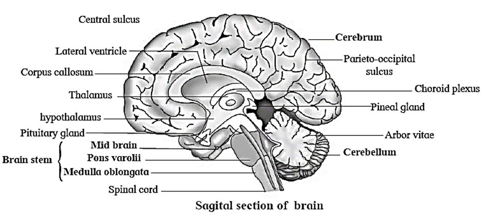
Forebrain : Forebrain consists of olfactory lobes, cerebrum and diencephalon.
Parts of the Forebrain (Prosencephalon) & functions. (i) Olfactory lobes (Rhinencephalon) (ii) Cerebrum (Telencephalon) 85% of brain Structure associated : Peculiarities : Functions of cerebrum : (iii) Diencephalon (Thalamencephalon) : Structure associated : Peculiarities : Functions of diencephalon :
Mid brain : It is located between diencephalon and the pons varolli. It contains the cerebral aqueduct or iter that connects the third and fourth ventricles.
Parts of the Midbrain (Mesencephalon) & Functions : (i) Corpora quadrigemina : Structure associated : Peculiarities : Functions of Corpora quadrigemina :
Hind brain : The posterior region of the brain is called hind brain.
Parts of the Hindbrain (Rhombencephalon) & Functions : (i) Cerebellum (Metencephalon) : 11% of the brain second largest region. Structure associated : Peculiarities : Functions of Cerebellum : (ii) Pons Varolii : Functions of Pons : (iii) Medulla oblongata (Myelencephalon) : Functions of Medulla oblongata :
Ventricles of brain : Ventricles are the cavities present in different parts of the brain.

Important terms associated with brain. Corpus callosum : Transverse band of nerve fibres which connects right and left cerebral hemisphere. It is the largest commissure of the brain. Cerebral cortex : The outer surface of cerebrum. composed of grey matter. Cerebral medulla : Inner part composed of white matter. Gyri (elevations) and Sulci (depressions) : convolutions and grooves on the surface of cerebrum. Central sulcus : Between frontal lobe and the parietal lobes. Parieto-occipital sulcus : Between parietal and occipital lobes. Lateral or Sylvian sulcus : Between temporal lobe and frontal and parietal lobes. Insula or insular cortex : Fifth lobe which is folded deep within the lateral sulcus. Foramen of Monroe : Narrow opening through which two lateral ventricles communicate with diocoel (third ventricle). Pineal gland : Vestigial 3rd eye and an important endocrine gland, producing hormones melatonin and serotonin. Habenular commissure : Connects two thalami. RAS (Reticular Activating System) : Relay centre as it transmits all sensory impulses except those of olfactory to the cerebrum.Situated in thalami. Aqueduct of Sylvius or iter : Connection between third and fourth ventricle through hypothalamus and midbrain. Limbic system : A complex neuronal circuit formed by the hypothalamus, amygdala, parts of epithalamus and thalamus, hippocampus and other areas. Optic chiasma : Crossing of the two optic nerves. Corpora quadrigemina : Four rounded elevations on the dorsal surface of the midbrain. The two superior colliculi are involved in visual reflexes and the two inferior colliculi are for auditory reflexes. Crura cerebri : Two thick fibrous tracks, also called cerebral peduncles, situated in the floor midbrain. Red nucleus : Grey matter near the centre of the midbrain, controlling posture and muscle tone, modifying some motor activities and motor coordination. Pons varolii : Rounded bulge on the underside of the brain stem. Brain stem : Consist of midbrain, pons and medulla. Arbor vitae : The mixing of white matter with the grey matter showing‘ a branched tree-like pattern. Cerebellar peduncles : Three pairs of myelinated nerve bundles connecting cerebellum to the other parts of CNS. A pair of lateral—foramina of Luschka and a median - foramen of Magendie: apertures on the posterior choroid plexus.
Spinal Cord : Spinal cord is the part of central nervous system and forms the lower extension of the medulla oblongata of the brain.
- It lies within the neural canal of the vertebral column and is surrounded by three meninges.
- Externally, the spinal cord appears as long cylindrical rod.
- It is 42 to 45 cm long and 2.0 to 2.5 cm broad.
- Conus medullaris : Terminal nervous part of the spinal cord.
- Filum terminale : Thread like non-nervous extension.
- 31 pairs of spinal nerves arise from lateral sides of the spinal cord.
- Cauda equina — Filum terminale with some spinal nerves running parallel to it. (appearing like a horse-tail)
T.S. of spinal cord : Functions :

Peripheral Nervous System (PNS) ; The peripheral nervous system connects the central nervous system to the different parts of the body having receptors and effectors.
Two types of peripheral nerves :
- Cranial nerves : arise from the brain.
- Spinal nerves : arise from the spinal cord.
Cranial nerves - nature and functions : diencephalon (Dentist’s nerve) a.Ophthalmic b. Maxillary c. Mandibular (largest) a.Sensory b.Sensory c. Mixed a. Nasal cavity, Upper eyelids, forehead, scalp, conjunctiva, lacrimal gland, scalp b. Mucosa of nose, palate, upper teeth, upper lip, lower eye lid parts of pharynx c. Lower teeth, skin over mandible cheek, side of head in front ear, muscles of mastication geniculate ganglion) neck muscles, lacrimal, sublingual, submandibula, nasal and palatine glands movement of neck, secretion of tears, taste, salivary secretion. cochlear) i. Vestibular ii. Cochlear - - movements like breathing cardiac, slowing, gastric and pancreatic secretion, gastrointestinal movements
Name
Type
Origin
Organs Innervated
Functions
1-Olfactory
Sensory
Olfactory bulb
Epithelium of Nose
Smell
2-Optic
Sensory
Side of
Retina of Eye
Vision
3-Occulomotor
Motor
Floor of mid brain
Eye muscles (4 of 6 eye muscles)
Movement of eye ball
4-Pathetic
Motor
Floor of mid brain
Eye muscles (1 of 6 eye muscles, forehead scalp)
Rotation and movement of eye ball
5-Trigeminal
Mixed
Ventral side of pons
-----
Sensation of skin touch, taste, jaw movement.
6-Abducens
Motor
Pons
Muscles of eye ball, lateral rectus muscle
Movement of eye
7-Facial (bearing
Mixed
Pons
facial, scalp and
Facial expression,
8-Auditory (vestibulo-
Sensory
Pons
Internal Ear
Hearing and equilibrium
9-Glossopharyngeal
Mixed
Side of medulla oblongata
Pharynx, tongue, salivary glands
Taste, salivation and swallowing
10-Vagus (Pneumogastric)
Mixed
Side of medulla oblongata
Larynx, trachea, pharynx, alimentary canal, heart, lungs, pancreas, blood vessels,
Visceral sensations and visceral
11-Spinal accessory
Motor
Side of medulla oblongata
Neck and shoulder muscles, reflexes of thoracic and abdominal vicera larynx, pharynx
Movements of larynx, pharynx, neck and shoulder
12-Hypoglossal
Motor
Side of medulla oblongata
Tongue muscles
Movement of tongue
Spinal Nerves :
- Thirty-one pairs of spinal nerves originate from the spinal cord.
- Spinal Nerves : All spinal nerves are mixed nerves.

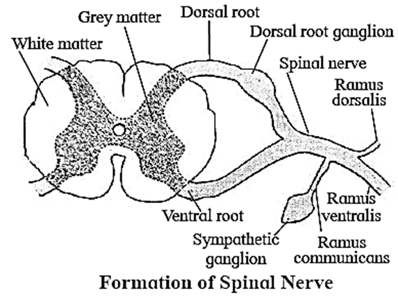
Formation of a typical spinal nerve :
- Each spinal nerve is formed inside the neural canal of vertebral column.
- The dorsal sensory and the ventral motor nerves together form the mixed spinal nerve.
As soon as it emerges out of vertebral column, it shows three branches, viz.
- Ramus dorsalis : from skin and to muscles of dorsal side
- Ramus ventralis : the largest of the three supplies the organs and muscles on lateral and anterior side
- Ramus communicans : the smallest of the three and given out from 1st thoracic upto 3rd lumbar (L3) spinal nerve. It joins the sympathetic ganglia.
Reflex Action :
Reflex action is defined as a quick, automatic involuntary and often unconscious action brought about when the receptors are stimulated by external or internal stimuli.
The path along which the action is carried out is called reflex arc.
Reflex arc : Reflex actions are controlled by CNS. Reflex arc is the structural or functional unit of reflex action. Simple reflex arc is formed of the following five components.
Types of reflexes : Based on the location of their action : The reflexes are divided into somatic reflexes and visceral reflexes. Based on the basis of number of neurons : Reflexes are of two types, viz. monosynaptic reflexes and polysynaptic reflexes. Based on inheritance and experience of learning: The reflexes are subdivided into unconditional or inborn and conditional or acquired. On the basis of control over the actions :
According to recent studies, the ANS is under the control of CNS and nerves arising from it (PNS).
According to this view, the PNS is divided into
- Somatic nervous system
- Autonomic nervous system
The somatic nervous system relays impulses from CNS to the skeletal or voluntary muscles of the body.
Autonomic Nervous System (ANS) : Autonomic nervous system transmits impulses from CNS to the involuntary organs and smooth muscles of the body.
It includes — autonomic ganglia, preganglionic fibres and postganglionic fibres.
Autonomic ganglia include
- Sympathetic ganglia-present near CNS in the form of sympathetic cord.
- Parasympathetic ganglia - present near or on the effector organs.
Preganglionic fibres arise from grey matter of CNS and end at autonomic ganglia.
Postganglionic fibres arise from autonomic ganglia to the effector organs.
Autonomic nervous system consists of sympathetic and parasympathetic nervous system.
(i) Sympathetic Nervous System [SNS) : (ii) Parasympathetic Nervous System : Comparison between Sympathetic and Parasympathetic Nervous System : (stomach and intestine)
Organ/Region
Sympathetic effect
Parasympathetic effect
Heart beat
Increases
Decreases
Blood vessels
Constricts
Dilates
Arterial B.P.
Increases
Decreases
Pupil of Eye
Dilates
Constricts
Gastrointestinal movements
Retards peristalsis
Accelerates peristalsis
Urinary bladder
Relaxes the bladder
Contracts the bladder
Sensory Receptors
Specialised structures in the body modified to receive the various stimuli from the external or internal environment.
Classification of receptors : Receptors are classified on the basis of their location, function and their sensitivity to specific stimuli. Their classification is given in the following chart.
Types of exteroceptors and interoceptors, their locations and functions : • Gustatoreceptors • Olfactory receptors Taste buds of tongue Olfactory Epithelium of Nose (*These are also considered as mechanoreceptors, receiving signals from internal organ)
No.
Name/Type of receptor
Location
Function
I. Exteroceptors : Receive external stimuli
a. Phonoreceptors
Internal Ear - organ of corti
Sound reception
b. Statoreceptors
Internal Ear- semicircular canals
Receptors for maintaining balance and equilibrium
c. Photoreceptors
Retina of Eye
Receives sensory stimuli for vision
d. Thermoreceptors
Skin
Receives sensory stimuli for heat (caloriceptors) and cold (trigidocetptors)
e. Mechanoreceptors
Skin
Sensitive to mechanical stimuli like touch, pain, pressure, deep pressure, etc.
f. Chemoreceptors
Sensitive to taste of sweet, salt, sour, bitter and umami. Sensitive to about 10,000 different smells
II. Interoceptors : Receive stimuli coming from within the body
a. Enteroceptors
from internal body organs
Sensitive to stimuli coming from internal organs like hunger, thirst, pain, osmotic change
b. Proprioceptors
Joints, muscles and tendons
Detect changes in the movements of joints, tendons and muscles; pain, tension and sensitive to vibrations
c. Baroreceptors
Present in walls of atria, venae cavae, aortic arch, carotid sinus
Sense changes in B.P. so as to restore homeostasis through vasodilation or vasoconstriction
Eye :
- The eyes are a pair of sensory organs of vision located in the orbit of skull
- Each eye is spherical/rounded and called eyeball.
Wall of the eyeball is made up of 3 layers : (1) sclera, (2) choroid (3) retina. Sclera is the outer layer of dense connective tissue with anterior transparent cornea. Choroid is the middle layer. It is bluish in colour containing many blood vessels. The anterior region is thick and forms the ciliary body. Posterior 2/3rd region is thinner. Iris is the forward segment of the ciliary body which is pigmented and opaque. This part is the visible coloured portion of the eye. Lens is present anteriorly inside the iris and is held in position by the ligaments of ciliary body. The aperture surrounded by the iris in front of the lens is known as pupil. The movement of the pupil is regulated by the muscle fibres of iris. Retina : The innermost layer of the eye is the retina having three sub-layers formed by ganglion cells, bipolar cells and photoreceptor cells, which are sensitive to light. Photoreceptor cells : There are two types of photoreceptor cells, viz. rods and cones containing light sensitive proteins. They are termed as photo pigments, rhodopsin which is a derivative of vitamin A (in rods) and iodopsin (in cones). Fovea is a central pit present beside it. Fovea is a thinned out portion of the retina where only the cones are densely packed and therefore have greatest visual acuity (resolution). A space between the cornea and the lens is called aqueous chamber. It contains a thin watery fluid known as aqueous humor.


Generation of image : Mechanism of vision :
1-Nerve impulse is transmitted by optic nerve to brain ↓
2-Nerve impulse in the axons of ganglion cells converge and leave via the optic nerve ↓
3-Nerve impulse transmitted to ganglion cells ↓
4-Nerve impulse transmitted to bipolar nerve cells ↓
5-Stimulation of rod and cone cells and generation of nerve impulse ↓
6-Breaking up of light sensitive pigments by specific wavelength of light ↓
7-Light falls on rod and cone cells in Retina ↓
8-Changes in retina when light rays fall on it ↓
9-Perception of image by the brain cells (in the visual area of cerebrum) ↓
Ear :
The human ear is called statoacoustic organ as it has two functions - hearing and body equilibrium
Anatomically the ear is made up of three divisions : the external ear, middle ear and inner ear,
External ear : It consists of ear pinna, auditory canal and tympanic membrane. Middle ear : It consists of chain of three ossicles called malleus, incus and stapes. Internal ear : It is fluid filled structure called labyrinth. It has two parts, bony and the membranous labyrinth.


Mechanism of Hearing :
Disorders of nervous system : Psychological disorders : Commonly called mental disorders. There is a wide range of conditions that affect the mood, thinking or behaviour. Some of the major categories of psychological disorders are : Parkinson’s disease : Alzheimer’s disease : The tympanic membrane (ear drum) is a delicate, membranous structure which transmits the sound waves to the middle ear.
Sir this pdf not download
Please download option i request you sir
Click on Buy button.