|
Topics to be Learn :
- Cell
- Kinds of Cells
- Components of Eukaryotic Cells
|
Cell : Cell is defined as a structural and functional unit of life of all living organisms capable of independent existence and can perform all functions of life.
- There is no typical shape of a cell. Cells may be spherical, rectangular, flattened, polygonal, oval, triangular, conical, columnar, etc.
- Smallest cell : Mycoplasma (0.3pm)
- Longest cell in animals : Nerve cell
- Largest cell : Ostrich egg
- Cell walls were first observed by Robertrly-Iooke (1665) as he looked through a microscope at dead cells from the bark of an oak tree. But Anton van Leeuwenhoek was first to visualize living cells using a single-lens microscope of his own construction.
- The Dutch scientist Anton van'Leeuwenh0ek made microscopes capable of magnifying single-celled organisms in a drop of pond water.
Instrument used for observing smaller organisms or cells :
Instrument used for observing smaller organisms or cells :
- To observe cells or organisms of smaller size we use a microscope.
- Larger cells can be seen through simple microscope but to observe smaller cells we require compound microscope. .
- Simple microscope can magnify image 50 to 100 times but a compound microscope can do so 1000 times or more. ‘
- In the microscope a beam of light is used to make things visible hence it is light microscope.
- To observe interior of cell we need electron microscope which can magnify image 500000 times.
[collapse]
Totipotency : Totipotency (totus : entire, potential: power) is the capacity or the potential of living nucleated cell, to differentiate into any other type of cell and thus, can form a complete new organism.
- A cell is totipotent because it has the entire genetic information of the organism in its nucleus.
- Embryonic animal cells are totipotent and are termed as stem cells.
- Stem cells are used in curing many diseases. Therefore, they have great potential for medical applications.
Cell theory : Schwann and Schleiden proposed the cell theory.
Postulates of modern cell theory: :
Postulates of modern cell theory:
- All living organisms are made up of cells.
- Living cells arise from pre-existing cells.
- A cell is the structural and functional unit of life.
- Total activities of cells are responsible for activity of an organism.
- Cells show transformation of energy.
- Cells contain nucleic acids; DNA and RNA in the nucleus and cytoplasm.
[collapse]
Kinds of Cells :
Categories of living organisms : Living organisms are grouped into two main categories the Prokaryotes and Eukaryotes.
Prokaryotic cells :
Characteristics of prokaryotic cell :
Characteristics of prokaryotic cell :
- Prokaryotic cells are primitive type of cells.
- It does not have membrane bound cell organelles (like endoplasmic reticulum, Golgi complex, mltochondrlai etc.) and well-defined nucleus (nuclear membrane is absent).
- Genetic material is in the form of nucleoid.

The cell in prokaryotes show following main features :
Cell envelope:
- Prokaryotic cell has chemically complex protective cell envelope having glycocalyx, cell wall and plasma membrane.
- In some bacteria, glycocalyx occurs in the form of a slime layer (loose sheath). Other bacteria may have a thick and tough covering called capsule. It helps in protection of bacterial cell.
Cell wall:
- The Gram-positive bacteria show presence of peptidoglycan layer in the cell wall and Gram-negative bacteria show presence of murein in the cell wall. It gives mechanical strength to the cell.
- In Gram-negative bacteria, cell wall is made up of two layers; inner layer of Murein or peptidoglycan and outer layer of Lipopolysaccharides.
Cell membrane:
- It is the innermost covering of the cell envelope, chemically composed of lipids and proteins.
- It helps in intercellular communication.
- Cell membrane shows infoldings called mesosomes which help in cell wall formation and DNA replication.
- The cyanobacteria show more longer extensions called as chromatophores which carry photosynthetic pigments.
In motile bacteria either cilia or flagella are found. Both are driven by rotatory movement produced by basal body (which works as motor) of flagellum. Other parts of flagellum are filament and hook.
Some other surface projections are the tubular pili (which help in inter-cellular communication) and fimbriae (for clinging to support).
[collapse]
Ribosomes:
- Bacterial cell cytoplasm contains dense particles called ribosomes which help in protein synthesis.
- Ribosomes are 70S type (composed of a larger sub-unit SOS and + smaller sub-unit 30S).
Q. Why bacterial nuéleus is said to be primitive?
Answer :
The DNA-containing central region of bacterial nucleus (prokaryotic cells) i.e. nucleoid, has no nuclear membrane separating it from the cytoplasm. Therefore, bacterial nucleus is said to be primitive.
[collapse]
Gram staining : The technique used for differentiating bacterial cells is
Gram staining.
Two types of bacteria, Gram—positive and Gram-negative can be identified using a staining process called the Gram staining,
Difference between Gram-positive and Gram-negative bacterial cells :
Difference between Gram-positive and Gram-negative bacterial cells :
- The Gram-positive bacteria show presence of peptidoglycan layer in the cell wall and Gram-negative bacteria show presence of murein in the cell wall.
- Gram-positive bacteria have a thicker peptidoglycan wall and stain a purple color, whereas the Gram-negative bacteria contain less peptidoglycan and do not retain the purple-colored dye.
[collapse]
Prokaryotic cytoplasm :
- Cytoplasm of prokaryotes is a pool of all necessary materials like water, enzymes, elements, amino acids, etc.
- Some inclusion bodies in form of organic (cyanophycean starch and glycogen) and inorganic granules (phosphate and sulphur) are also found.
Bacterial flagellum : The bacterial flagellum is an organelle for motility made up of three parts:
- The basal body that spans the cell envelope and works as_ a rotary motor;
- The helical filament that acts as a propeller;
- The hook that acts as a universal joint connecting these two to transmit motor torque to the propeller.
The motor i.e. basal body drives the rotation of the long, helical filamentous propeller at hundreds of hertz to produce thrust that allows bacteria to swim in liquid environments.
Therefore, basal body of bacterial flagella considered as smallest motor in the world.
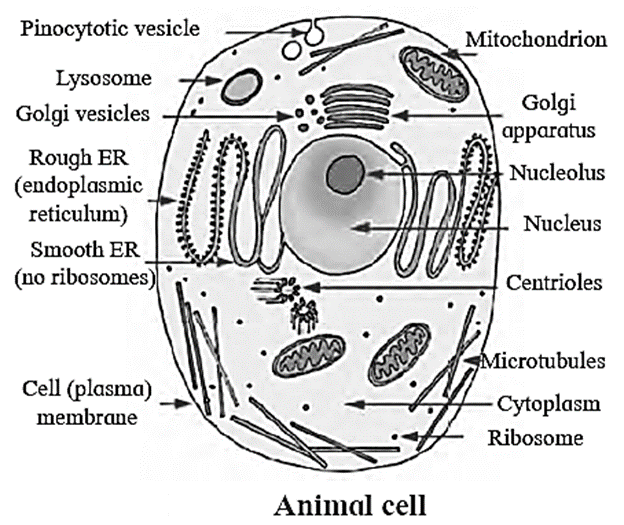
Eukaryotic cells : Eukaryotic cells are the cells possessing well-defined nucleus and membrane bound organelles
- Examples: mitochondria, endoplasmic reticulum, ribosomes, Golgi complex etc.
- Eukaryotes include protists, plants, animals and fungi.
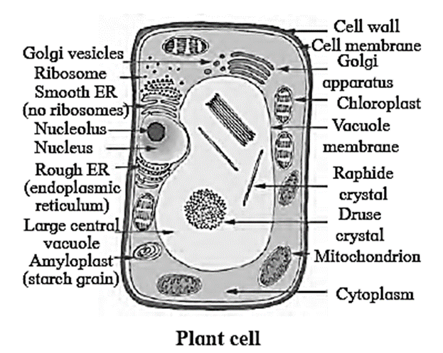
Cell wall in Eukaryotic cells :
Cell wall in Eukaryotic cells : The rigid, protective and supportive covering, outside the cell membrane is called cell wall.
- It is present in plant cells, fungi and some protists.
- Algae show presence of cellulose, galactans, mannans and minerals like calcium carbonate in cell wall.
- In other plants, it is made up of hemicelluloses, pectin, lipids and protein.
- Microfibrils of plant cell wall show presence of cellulose which is responsible for rigidity.
- Some of the depositions of cell wall are silica (grass stem), cutin (epidermal walls of land plants), suberin (endodermal cells of root), wax, lignin.
Function:
- Provides support, rigidity and shape to the cell.
- Protects the protoplasm against mechanical injury and infections.
[collapse]
Differences between prokaryotic and eukaryotic cells :
Differences between prokaryotic and eukaryotic cells :
| Prokaryotic cell |
Eukaryotic cell |
| 1-Nuclear membrane is absent.
2-Genetic material is in the form of circular coil of DNA without histone proteins.
3-Membrane bound cell organelles are absent.
4-Plasmids are many in number,
5-Cytoplasm does not show streaming movement.
6-Ribosomes are smaller and of 70S type.
7-Respiratory enzymes are present on the infoldings of the plasma membrane called mesosomes.
8- Ex. Cyanobacteria (Blue green algae) and Bacteria.
|
1-Nuclear membrane is present.
2-Genetic material is in the form of a double helix DNA with histone proteins,
3-Membrane bound cell organelles are present.
4-Plasmids are absent,
5-Cytoplasm shows streaming movement.
6-Ribosomes are larger and of 80S type.
7-Respiratory enzymes are present within
8-Ex. Algae, fungi, plants and animals.
|
[collapse]
Plant cell :
- Plant cells show presence of plasmodesmata which are cytoplasmic bridges between neighbouring cells.
- This open channel through the cell wall connects the cytoplasm of adjacent plant cells and allows water, small solutes, and some larger molecules to pass between the cells. In this wav though plants have no circulatory system, plant cells manage intercellular transport.
Structure of plant cell wall :
Structure of plant cell wall :
In plants, cell wall shows middle lamella, primary wall and secondary wall
- Middle lamella : It is thin and present between two adjacent cells. It is the first structure formed from cell plate during cytokinesis. It is mainly made up of pectin, calcium and magnesium pectate. Softening of ripe fruit is due to solubilization of pectin.
- Primary wall: In young plant cell, it is capable of growth. It is laid inside to middle lamella. It is the only wall seen in meristematic tissue, mesophyll, pith, etc.
- Secondary wall: It is present inner to primary wall. Once the growth of primary wall stops, secondary wall is laid. At some places thickening is absent which leads to formation of pits.
[collapse]
Eukaryotic plasma membrane/ Cell membrane/ Bio-membrane :
Eukaryotic plasma membrane/ Cell membrane/ Bio-membrane :
- It is thin, quasi-fluid structure present both extracellularly and intracellularly.
- Extracellularly, it is present around protoplast and intracellularly, it is present around most of the cell organelles in eukaryotic cell. It separates cell organelles from cytosol.
- Thickness of bio-membrane is about 75A.
- Cell membrane appears trilaminar (made up of three layers) when observed under electron microscope. It shows presence of lipids (mostly phospholipids) arranged in bilayer.
- Lipids possess one hydrophilic polar head and two hydrophobic non-polar tails. Therefore, phospholipids are amphipathic.
- Lipid molecules are arranged in two layers (bilayer) in such a way that their tails are sandwiched in between heads. Due to this, tails never come in direct contact with aqueous surrounding.
- Cell membrane also shows presence of proteins and carbohydrates.
- Ratio of proteins and lipids varies in different cells. For example, in human beings, RBCs show approximately 52% protein and 40% lipids.
[collapse]
Fluid mosaic model :
Fluid mosaic model : It is most accepted model of cell membrane. It was proposed by Singer and Nicholson in 1972.
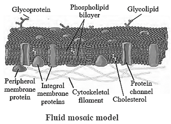
- This model states that plasma membrane is made up of phospholipid bilayer and proteins.
- Proteins are embedded in the lipid membrane like icebergs in the sea of lipids.
- Phospholipid bilayer is fluid in nature.
- Quasi-fluid nature of lipid enables lateral movement of proteins. This ability to move within the membrane is measured as fluidity.
Based on organization of membrane proteins they are of two types, as:
- A. The intrinsic proteins occur at different depths of bilayer i.e. they are tightly bound to the phospholipid bilayer and are embedded in it. They span the entire thickness of the membrane. Therefore, they are known as transmembrane proteins. They form channels for passage of water.
- B. The extrinsic or peripheral proteins are found on two surfaces of the membrane i.e. are loosely held to the phospholipid layer and can be easily removed.
[collapse]
| Glycoproteins : Glycoproteins are protein molecules modified within the Golgi complex by having a short sugar chain (polysaccharide) attached to them.
The polysaccharide part of glycoproteins located on the surfaces of red blood cells acts as the antigen responsible for determining the blood group of an individual. .
Different polysaccharide part of glycoproteins act as different type of antigens that determine the blood groups. ,
Four types of blood groups -A, B, AB, and O are recognized on the basis of presence or absence of these antigens.
|
Functions of plasma membrane :
Functions of plasma membrane : The significant function of plasma membrane is transport of molecules across it. Plasma membrane is selectively permeable.
- Passive transport: During passive transport, many molecules move across the membrane without spending energy. Some solutes move by simple diffusion along the concentration gradient (from higher to lower concentration). Neutral solutes may move across the membrane by the process of simple diffusion This is called the passive transport. Water may also move by osmosis.-
- Active transport: During active transport, few ions or molecules are transported against concentration gradient i.e. from lower to higher concentration. This requires energy, hence ATP is utilized. As such a transport is an energy dependent process in which ATP is utilized, it is called Active transport e. g. Na+/K+ pump. Polar molecules cannot pass through non-polar lipid bilayer. Therefore, they require carrier proteins to facilitate their transport across the membrane.
- Plasma membrane imparts shape to the cell and also protects the cell from injury.
- Plasma membrane regulates the cellular semi-permeability, reabsorption, excretion and secretion.
- Various cell organelles can be formed within the cell, with the help of plasma membrane.
- It regulates cellular interactions in the formation of tissues and the defence against foreign bodies.
- The cell membrane separates protoplasm from the external environment to maintain the individuality of the cell.
- It acts as a receptor for various chemical stimuli such as amino acids, hormones and sugars.
- In unicellular organisms like Amoeba, pseudopodia are formed by projection of plasma membrane.
- Ingestion of food and water by endocytosis or pinocytosis takes place with the help of plasma membrane.
[collapse]
Cytoplasm in Eukaryotic cell :
- The cell contains ground substance called cytoplasmic matrix or cytosol.
- This colloidal jelly like material shows streaming movements called cyclosis.
- The cytoplasm contains water as major component along with organic and inorganic molecules like sugars amino acids, vitamins, enzymes, nucleotides, minerals and waste products.
- It also contains various membrane bound cell organelles like endoplasmic reticulum, Golgi complex, mitochondria, plastids, nucleus, microbodies and cytoskeletal elements like microtubules.
- Cytoplasm acts as a source of raw materials as well as seat for various metabolic activities taking place in the cell.
- It helps in distribution and exchange of materials between various cell organelles.
Endomembrane system of the cell :
- Cell organelles are compartments in the cell that carry out specific functions.
- Some of these organelles coordinate with each other and complete the specific function of the cell.
- Nuclear membrane, endoplasmic reticulum, Golgi complex, lysosomes and various types of vesicles and vacuoles form such a group and are together considered as endomembrane system of the cell.
Q. Why mitochondria and chloroplasts are not considered as a part of endomembrane system?
Answer :
Ans. Organelles having distinct functions are not included in endomembrane system.
Mitochondria or chloroplast carry out specific type of energy conversions in the cell.
Therefore, mitochondria and chloroplasts are not considered as a part of endomembrane system.
[collapse]
Structure of Endoplasmic Reticulum :
Structure of Endoplasmic Reticulum :
- Endoplasmic reticulum is a network present within the cytosol.
- It is present in all eukaryotic cells except ova and mature red blood corpuscles.
- Under the electron microscope, it appears like network of membranous tubules and sacs called cisternae.
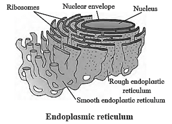
- This network of ER divides the cytoplasm in two parts viz. one within the lumen of ER called laminal cytoplasm and non-laminal cytoplasm that lies outside ER.
- Membrane of ER is continuous with nuclear envelope at one end and extends till cell membrane. It thus acts as intracellular supporting framework and helps in maintaining position of various cell organelles in the cytoplasm.
- Depending upon the presence or absence of ribosomes, endoplasmic reticulum is called rough endoplasmic reticulum (RER) or smooth endoplasmic reticulum (SER) respectively.
[collapse]
Smooth endoplasmic reticulum (SER):
- Depending on cell type, it helps in synthesis of lipids for e.g. Steroid secreting cells of cortical region of adrenal gland, testes and ovaries.
- Smooth endoplasmic reticulum plays a role in detoxification in the liver and storage of calcium ions (muscle cells).
Rough Endoplasmic Reticulum (RER):
- Rough ER is primarily involved in protein synthesis. For e.g. Pancreatic cells synthesize the protein insulin in the ER.
- These proteins are secreted by ribosomes attached to rough ER and are called secretory proteins. These proteins get wrapped in membrane that buds off from transitional region of ER. Such membrane bound proteins depart from ER as transport vesicles
- Rough ER is also involved in formation of membrane for the cell. The ER membrane grows in place by addition of membrane proteins and phospholipids to its own membrane. Portions of this expanded membrane are transferred to other components of endomembrane system.
Golgi complex : Golgi complex or Golgi apparatus or Golgi body act as a assembly, manufacturing cum packaging and transport unit of cell.
Structure of Golgi complex :
Structure of Golgi complex:
- Golgi complex consists of stacks of membranous sacs called cisternae.
- Diameter of cisternae varies from 0.5 to 1 um.
- A Golgi complex may have few to several cisternae depending on its function.
- The thickness and molecular composition of membranes at one end of the stack of a Golgi sac differ from those at the other end.
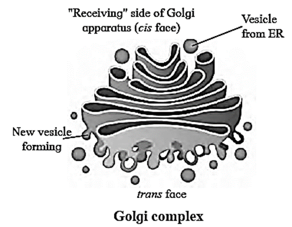
- The Golgi sacs show specific orientation in the cell.
- Each cisterna has a forming or ‘cis’ face (cis: on the same side) and maturing or ‘trans’ face (trans: the opposite side).
- Transport vesicles that pinch off from transitional ER merge with cis face of Golgi cisterna and add its contents into the lumen.
- While transport vesicles are leaving from the trans face of the Golgi, certain markers get impregnated on their membrane. These markers help them to identify their specific target cell or cell organelle.
[collapse]
The cisternae in Golgi body are not physically connected to each other as that are in ER.
According to recent studies it is proposed that cisternae of Golgi body themselves mature moving from cis to trans face. It is called ‘Cisternal maturation model’.
It is also said that some vesicles recycle their enzymes that have been carried forward by moving cisternae back to less mature region.
Location of Golgi complex:
- Golgi bodies are usually located near endoplasmic reticulum.
Functions of Golgi complex: :
Functions of Golgi complex:
- Golgi body carries out two types of functions, modification of secretions of ER and production of its own secretions.
- Cisternae contain specific enzymes for specific functions.
- Refining (modification) of product takes place in a sequential manner.
- For example, certain sugar component is added or removed from glycolipids and glycoproteins that are brought from ER, thus forming a variety of products.
- Golgi bodies also manufacture their own products. Golgi bodies in many plant cells produce non-cellulose polysaccharides like pectin.
- Manufactured or modified, all products of Golgi complex leave cisternae from trans face as transport vesicles.
[collapse]
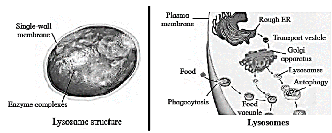
Lysosomes :
- Lysosomes are considered as dismantling and restructuring units of a cell.
- These are membrane bound vesicles containing hydrolytic enzymes. The enzymes in lysosomes are used by most eukaryotic cells to digest (hydrolyse) macromolecules.
- The lysosomal enzymes show optimal activity in acidic pH.
- Lysosomes arise from Golgi associated endoplasmic reticulum.
- Lysosomes are polymorphic in nature and are classified as primary lysosomes, secondary or hybrid lysosomes, residual body and autophagic vesicle. -
- The list of lysosomal enzymes includes: - All types of hydrolases viz, amylases, proteases and lipases.
Characteristics of lysosomes :
Characteristics of lysosomes :
Lysosomes are polymorphic in nature.
- Lysosomes are classified as, Primary lysosomes; which are nothing but membrane bound vesicles in which enzymes are in inactive state.
- Secondary lysosomes or hybrid lysosomes, which are formed by fusion of lysosome with endocytic vesicle containing materials to be digested, represented as heterophagic vesicle. This is larger in size than primary lysosome.
- When organic molecules or membrane bound old cell organelle to be recycled fuses with primary lysosome, autophagic vesicles are formed.
- Residual body is the vesicle containing undigested remains left over in the heterophagic vesicle after releasing the products of digestion in the cytosol.
- 'Hence, lysosomes are polymorphic in nature.
Lysosomes are called suicide bags of the cells.
- Lysosomes which bring about digestion of cell’s own organic material like a damaged cell organelle are called autophagic vesicle (suicide bags).
- An autophagic vesicle essentially consists‘ of lysosome fused with membrane bound old cell organelle or organic molecules to be recycled.
- Thus, lysosomes are capable of destructing all kinds of material in the cell. Therefore, can digest its own cell organelles due to presence of lysosome.
- Hence, lysosomes are also called as suicide bags.
[collapse]
Intracellular and Extracellular digestion :
Intracellular digestion:
- The intracellular digestion is brought about by autophagic vesicle or secondary lysosomes which contain foreign materials brought in by processes like phagocytosis.
- E. g. Food vacuole in amoeba oi" macrophages in human blood that engulf and destroy harmful microbes that enter the body.
Extracellular digestion:
- Extracellular digestion is brought about by release of lysosomal enzymes outside the cell.
- E.g. acrosome a cap like structure in human sperm is a modified lysosome which contain various enzymes like Hyaluronidase. These enzymes bring about fertilization by dissolving protective layers of ovum.
[collapse]
Vacuole : The organelle which helps in maintaining turgidity of the cell and a proper internal balance of cellular contents is known as vacuole.
- The vacuoles are bound by semipermeable membrane, called tonoplast membrane. This membrane helps in maintaining the composition of vacuolar fluid (cell sap), different from that of the cytosol.
- Composition of cell sap differs in different types of cells.
- Vacuoles store excretory products or even compounds that are harmful or unpalatable to herbivores, thereby protecting the plants.
- Attractive colours of the petals are due to storage of such pigments in vacuoles.
- Generally, there are two or three permanent vacuoles in a plant cell. -
- In some large plant cells, a single large vacuole occupies the central part of the cell. It is called central vacuole. In such cells, vacuole can occupy about 90% of the total volume of the cell.
- The cell sap of central vacuole is a store house of various ions and thus is hypertonic to cytosol.
- Small vacuoles in seeds of certain plants store organic materials like proteins.
- In animal cells, they are few in number and smaller in size.
- Intake of food or foreign particle by phagocytosis involves formation of food vacuole.
Function of contractile vacuole in Paramoecium :
Contractile vacuole perform excretion and osmoregulation in fresh water unicellular forms like Paramoecium.
Microbodies : Microbodies are minute membrane bound sacs found in both plant and animal cells.
Types and functions of Microbodies :
Types and functions of Microbodies :
Microbodies contain various types of enzymes based on which they are classified into following types:
Sphaerosomes:.
- These are found mainly in cells involved in synthesis and storage of fats. For e. g. endosperm of oil seeds.
- The membrane of sphaerosome is half unit membrane i.e. this membrane has only one phospholipid layer.
Peroxisomes :
- Peroxisomes contain enzymes that remove hydrogen atoms from substrate and produce toxic hydrogen peroxide by utilisation of oxygen.
- At the same time peroxisome also contains enzymes that convert toxic H202 to water. Conversion of toxic substances like alcohol takes place in liver cells by peroxisomes.
[collapse]
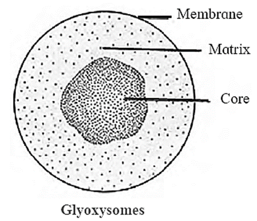
Glyoxysomes : Glyoxysomes are membrane bound organelles containing enzymes that convert fatty acids to sugar. They are observed in cells of germinating seeds where the cells utilize sugar (formed by conversion of stored fatty acids) till it starts photosynthesising on its own.
Mitochondrion : These are important cell organelles involved in aerobic respiration.
Mitochondrion is known as the power house of the cell. Mitochondria are absent in prokaryotic cells and red blood corpuscles (RBCs).
Structure of mitochondrion :
The structure of mitochondrion :
- Shape of the mitochondria may be oval or spherical or like spiral strip.
- It is a double membrane bound organelle.
- Outer membrane is permeable to various metabolites due to presence of a protein-Porin or Parson’s particles.
- Inner membrane is selectively permeable to few substances only.
- Both membranes are separated by intermembrane space.
- Inner membrane shows several finger like or plate like folds called as cristae which bears numerous particles oxysomes and cytochromes / electron carriers.
- Inner membrane encloses a cavity called inner chamber, containing a fluid-matrix.
- Matrix contains few coils of circular DNA, RNA, 70S types of ribosomes, lipids and various enzymes of Krebs’ cycle and other pathways.
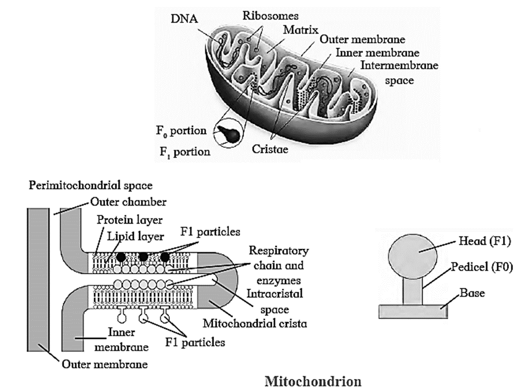
[collapse]
Location of mitochondria :
- Mitochondria are found in nearly all eukaryotic cells, including plants, animals, fungi, and most unicellular eukaryotes.
- Some of the cells have a single large mitochondrion, but frequently a cell has hundreds of mitochondria.
- The number of mitochondria correlates with the cell’s level of metabolic activity. For e.g. cells that move or contract have proportionally more mitochondria than metabolically less active cells.
- However, mature red blood cells in humans lack mitochondria.
Oxysome/ F1 Particle. :
Oxysome/ F1 Particle.
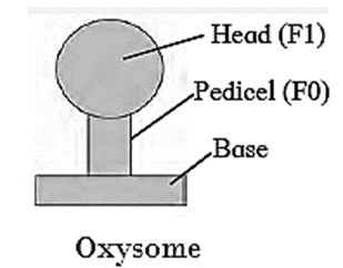
Structure of Oxysome :
- Inner membrane of mitochondria bears numerous particles called as Oxysomes (F1-F0 / Fernandez-Moran Elementary particles / Mitochondrial particles).
- Each particle consists of head, stalk (Pedicel) and base.
- Head (F1) / lollipop head faces towards matrix and foot (F0) is embedded in inner membrane.
- Head acts as an enzyme ATP synthase and foot (base) as proton channel. Oxysomes are involved in proton pumping and ATP synthesis.
[collapse]
Plastids : Plastids are double walled organelles containing DNA, RNA and 70S ribosomes
Types of Plastids :
Types of Plastids :
Plastids are classified according to the pigments present in it. Three main types of plastids are — leucoplasts, chromoplasts and chloroplasts.
(i) Leucoplasts do not contain any photosynthetic pigments they are of various shapes and sizes. These are meant for storage of nutrients.
- Amyloplasts store starch.
- Elaioplasts store oils. .
- Aleuroplasts store proteins.
(ii) Chromoplasts contain pigments like carotene and xanthophyll etc.
- They impart yellow, orange or red colour to flowers and fruits.
- These plastids are found in the coloured parts of flowers and fruits.
(iii) Chloroplasts are plastids containing green pigment chlorophyll along with other enzymes that help in production of sugar by photosynthesis. They are present in plants, algae and few protists like Euglena.
[collapse]
Chloroplast : Chloroplast plays a significant role in synthesis of starch in plants.
Structure of chloroplast :
Structure of chloroplast:
- In plants, chloroplast is found mainly in mesophyll of leaf.
- Chloroplst is lens shaped but it can also be oval, spherical discoid or ribbon like.
- A cell may contain single large chloroplast as in Chlamydomonas or there can be 20 to 40 chloroplasts per cell as seen in mesophyll cells.
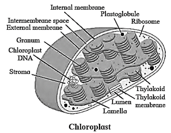
- Chloroplasts contain green pigment called chlorophyll along with other enzymes that help in production of sugar by photosynthesis. .
- Inner membrane of double walled chloroplast is comparatively less permeable.
- Inside the cavity of inner membrane, there is another set of membranous sacs called thylakoids.
- Thylakoids are arranged in the form of stacks called grana (singular: granum).
- The grana are connected to each other by means of membranous tubules called stroma lamellae.
- Space outside thylakoids is filled with stroma.
- The stroma and the space inside thylakoids contain various enzymes essential for photosynthesis.
- Stroma of chloroplast contains DNA and ribosomes (70S).
[collapse]
Ribosomes :
Ribosomes : Ribosomes are protein factories of the cell. They use the genetic information to synthesise proteins.

- Ribosomes were first observed as dense particles in electron micrograph of a cell by scientist Palade in 1953.
- Ribosomes lack membranous covering around them and are made up of Ribosomal RNA and proteins.
- In a eukaryotic cell, ribosomes are present in mitochondria, plastids (in plant cells) and in cytosol.
- Ribosomes are either found attached to outer surface of Rough Endoplasmic Reticulum and nuclear membrane or freely suspended in cytoplasm.
- Both are of 80S type. Each ribosome is made up of two subunits- a large (60S) and a small (408) subunit.
- Bound ribosomes generally produce proteins that are transported outside the cell after processing in ER and Golgi body. e.g. Bound ribosomes of acinar cells of pancreas produce pancreatic digestive enzymes.
- Free ribosomes come together and form chains called polyribosomes for protein synthesis.
- Free ribosomes generally produce enzymatic proteins that are used up in cytoplasm, like enzymes required for breakdown of sugar.
- Both types of ribosomes (bound and free) can interchange position and function.
- Number of ribosomes is high in cells actively engaged in protein synthesis.
The particle size of ribosomes is measured in terms of Svedberg unit (S). It is a measure of sedimentation rate of a particle in ultracentrifuge. It is thus a measure of density and size of a particle. 1S = 10-13 sec.
[collapse]
Nucleus : Nucleus is known as the master cell organelle as it regulates various metabolic activities through synthesis of various proteins and enzymes.
The nucleus in eukaryotic cell is made up of nuclear envelope, nucleoplasm, nucleolus and chromatin network.
Nuclear envelope :
Nuclear envelope :

- Nuclear envelope is a double walled delimiting membrane of nucleus.
- Two membranes are separated from each other by perinuclear space (10 to 50nm).
- Outer membrane is connected with endoplasmic reticulum at places and harbours ribosomes on it.
- The inner membrane is lined by nuclear lamina- a network of protein fibres that helps in maintaining shape of the nucleus.
- The two membranes along with perinuclear space help in "separating nucleoplasm from cytoplasm.
- However, nuclear membrane is not continuous.
- There are small openings called nucleopores on the nuclear membrane.
- The nucleopores are guarded by pore complexes which regulate flow of substances from nucleus to cytoplasm and in reverse direction.
[collapse]
Nucleoplasm or karyolymph :
- The nucleoplasm or karyolymph contains various substances like nucleic acids, protein molecules, minerals and salts.
- It contains chromatin network and nucleolus.
Nucleolus :
Nucleolus :

- Nucleolus is made up of rRNA and ribosomal proteins and it is known as the site of ribosome biogenesis.
- The rRNA and ribosomal proteins are transported to cytoplasm and are assembled together to form ribosomes.
- Depending on synthetic activity of a cell, thereare one or more nucleoli present in the nucleoplasm. For e.g. cells of oocyte contain large nucleolus whereas sperm cells contain small inconspicuous one.
- Nucleolus appear as dense spherical body present near chromatin network.
[collapse]
Functions of the nucleus:
- Nucleus is known as the controlling unit of-the cell.
- The nucleus contains entire genetic information, hence play important role in heredity and variation
- It is the site for synthesis of DNA, RNA and ribosomes.
- It plays important role in protein synthesis.
Chromatin material :
- Nucleus contains genetic information in the form of chromosomes which are DNA molecules associated with proteins.
- In a non-dividing cell, the chromosomes appear as thread like network and cannot be identified individually. This network is called chromatin material.
- The chromatin material contains DNA, histone and non-histone proteins and RNA.
- In some regions of chromatin, DNA is more and is genetically active called euchromatin.
- Some regions that contain more of proteins and less DNA and are genetically inert, are called heterochromatin
Heterochromatin is a region in chromatin that is highly compacted during interphase and is generally not accessible for transcription of genes.
Q. What is the significance of having constant chromosome number in species.
Answer :
Constant chromosome number in a species is important in phylogenetic studies.
[collapse]
Cytoskeleton : Presence of network of fibrils throughout the cytoplasm is called cytoskeleton.
- The cytoskeleton is a supportive structure built from microtubules, intermediate filaments, and microfilaments.
- Microtubules are made up of protein- tubulin. .
- Microfilaments are made up of actin.
- Intermediate filaments are composed of fibrous proteins.
Functions of Cytoskeleton :
Functions of Cytoskeleton :
- Maintenance of shape of cell,
- Contraction of cell,
- Mobility of cell and cell organelles,
- Changes in shape of the cells and cell division.
[collapse]
Cilia and flagella :
- They are fine hair like membrane bound protoplasmic outgrowths that occur on the free surface of the cell.
- Cilia are small in size and many in number.
- Cilia act as oars causing movement of cell.
- Flagella are longer and few in number.
- Flagella present in prokaryotic bacteria are structurally different from that of eukaryotic flagella.
Structure of Cilia/flagella :
Structure of Cilia/flagella :
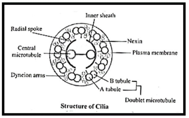
- Cilium or flagellum helps in locomotion of unicellular organisms.
- They consist of basal body, basal plate and shaft.
- Basal body is placed in outer part of cytoplasm. It is derived from centriole. It has nine peripheral triplets of fibrils.
- Shaft is exposed part of cilia or flagella. It consists of two parts sheath and axoneme.
- Sheath is covering membrane of cilium or flagellum.
- Core called axoneme possesses 11 fibrils (microtubules) running parallel to long axis.
- It shows 9 peripheral doublet microtubules and two single central microtubules (9+2).
- The central tubules are enclosed by central sheath.
- This sheath is connected to one of the tubules of peripheral doublets by a radial spoke.
- Central tubules are connected to each other by bridges
- The peripheral doublet microtubules are connected to each other through linkers or inter-doublet.
[collapse]
Centrioles and centrosomes :
Centrioles and centrosomes : Centrosome is usually found near the nucleus of an animal cell. It contains a pair of cylindrical structures called centrioles.

- Centrioles and centrosomes play significant role in formation of spindle apparatus during cell division.
- The cylinder (centriole) are perpendicular to each other and are surrounded by amorphous substance called pericentriolar material.
- Each cylinder of centriole is made up of nine sets of triplet microtubules made up of tubulin.
- Evenly spaced triplets are connected to each other by means of non-tubulin proteins.
- At the proximal end of centriole, there is a set of tubules called hub.
- The peripheral triplets are connected to hub by means of radial spokes. Due to this proximal end of centriole looks like a cartwheel.
- Centriole forms basal body of cilia and flagella.
[collapse]
It’s really amazing
Good organisation of information
It is very helpful for me. Thankyou so much.
That’s really helpful
good for exam….