|
Topics to be Learn :
- Heart
- Working mechanism of human heart
- Blood vessels
- Blood Pressure (BR)
- Electrocardiogram
- Lymphatic system
|
Heart :
- Heart is a hollow, muscular, conical organ about the size of one’s fist with broad base and narrow apex tilted towards the left.
- It is mesodermal in origin.
- It is situated in middle of the thoracic cavity in a space called mediastinum, between the two lungs.
- The heart is 12 cm in length, 9 cm in breadth and 250 to 300 grams in weight.
Pericardium : Double layered membrane, these layers are as follows :
- Fibrous pericardium : Outer, tough layer of inelastic fibrous connective tissue.
- Serous pericardium : This inner pericardium has two layers, outer parietal layer and inner visceral layer.
- Parietal layer forms the inner lining of fibrous pericardium.
- Visceral layer or epicardium is next to heart on the outer side.
- Pericardial fluid is present between the parietal and visceral layers of serous pericardium.
Heart wall :
- The heart wall has three layers, viz. outer epicardium, middle myocardium arid inner endocardium.
- Epicardium has single layer of flat epithelial cells called mesothelium.
- Myocardium has cardiac muscle fibres responsible for movements of the heart.
- Endocardium has single layer of flat epithelial cells called endothelium.

External structure of heart :
External structure of heart :
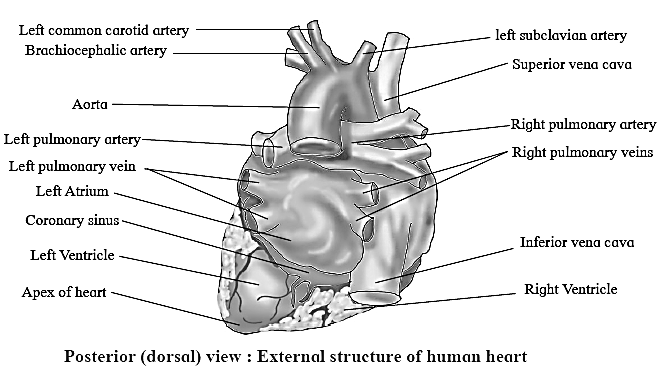
- Human heart consists of two superior, small, thin walled receiving chambers called atria or auricles and two inferior, large, thick walled, distributing chambers called ventricles.
- Atrio-ventricular groove or coronary sulcus, a transverse groove which is present between the atria and the ventricles is seen externally.
- The inter-ventricular sulcus is present between the right and left ventricles. Coronary arteries and coronary veins are present in the sulci. The coronary veins join to form coronary sinus which opens into the right atrium.
- The right atrium receives deoxygenated blood from all over the body through superior vena cava and inferior vena cava.
- Left atrium receives oxygenated blood from lungs through two pairs of pulmonary veins.
- From the right ventricle deoxygenerated blood is sent to lungs through pulmonary trunk.
- From the left ventricle oxygenated blood sent to entire body by systemic aorta.
- Ligamentum arteriosum connects the pulmonary trunk and systemic aorta. It represents ductus arteriosus of foetus.
[collapse]
Internal structure of heart :
Internal structure of heart :

- There are four chambers in the heart, viz, two atria and two ventricles which can be demarcated internally.
- Atria are thin walled upper receiving chambers separated from each other by interatrial septum.
- The right atrium receives deoxygenated blood from all over the body through superior vena cava, inferior vena cava and from the heart through coronary sinus.
- The opening of inferior vena cava is guarded by Eustachian valve while the opening of coronary sinus is guarded by Thebesian valve.
- The fossa ovalis is oval depression that is present on the right side of interatrial septum.
- It is the remnant of foramen ovale, an oval opening in the interatrial septum of the foetus.
- The left atrium receives oxygenated blood from the lungs through four openings of pulmonary veins.
- Right and left atria open into the right and left ventricles respectively through atrioventricular apertures. These are respectively guarded by tricuspid and bicuspid valves made up of connective tissue.
- The right atrioventricular valve has three flaps hence called tricuspid valve while left atrioventricular valve has two flaps hence called bicuspid valve or mitral valve.
- These valves are attached to papillary muscles of ventricles by chordae tendinae. The valves are prevented from turning back into the atria during the contraction of ventricles due to chordae tendinae.
- Ventricles are two thick walled lower, distributing chambers separated from each other by interventricular septum.
- Left ventricle has thick wall. The inner surface of the ventricle is thrown into a series of irregular muscular ridges called columnae carnae or trabeculae carnae.
- Pulmonary trunk or aorta arises from the right ventricle carrying deoxygenated blood to lungs for oxygenation. Systemic aorta arises from the left ventricle carrying oxygenated blood to all parts of the body.
- Pulmonary aorta and systemic aorta have three semilunar valves at the base which prevent the backward flow of blood during ventricular diastole.
[collapse]
Pumping action of heart: The heart acts as the main pumping organ of the circulatory system. The pumping action is brought about by a rhythmic contraction and relaxation of the cardiac muscles or heart muscles. Contraction of heart muscles is systole and relaxation of heart muscles is diastole.
- The rate of heartbeat is about 72 times per minute during which it pumps out about 5 litres of blood which equals cardiac output.
Conducting system of heart :
Conducting system of heart : The heartbeat in human beings originates in modified cardiac muscles called sinoatrial node (S.A. node). Therefore, the heart is said to be myogenic.

- The conducting system of heart consists of sinoatrial node (SAN), atrioventricular node (AVN), Bundle of His and Purkinje fibres.
- The heart shows auto-rhythmicity as the impulse for its rhythmic movement during beating is developed inside the heart.
- The autorhythmic fibres are developed during embryonic life. They act as pacemaker by setting the rhythm for the heart. They also form conducting system for conducting impulses throughout heart muscles.
- The impulse travels in the heart in the following manner : Sinoatrial node (Pacemaker) —> Internodal pathway —> Atrioventricular node —> Bundle of His —-> Right and left bundle branches —> Purkinje fibres.
[collapse]
Working mechanism of human heart :
Working mechanism of human heart :
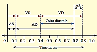
- Cardiac cycle : Human heart alternately contracts and relaxes. Contraction is called systole and relaxation is called diastole. Atria and ventricles contract alternately. Consecutive systole and diastole constitutes a single heartbeat or cardiac cycle. One atrial systole (0.1 second), one ventricular systole (0.3 second), followed by a joint diastole (0.4 second). One cardiac cycle takes place in about 0.8 second and is also called a heartbeat.
- Atrial systole : During atrial systole, the deoxygenated blood from the right atrium enters the right ventricle through atrioventricular aperture whereas the oxygenated blood from left atrium enters the left ventricle through atrioventricular aperture. In normal conditions atrial systole lasts for 0.1 second and atrial diastole lasts for 0.7 second.
- Ventricular systole : During ventricularsystole, the deoxygenated blood from the rightventricle enters the pulmonary trunk and theoxygenated blood from the left ventricle entersthe aorta. The backflow of blood into atria isprevented by the closure of cuspid valves ofboth atrioventricular apertures (lubb sound isproduced) Ventricular systole lasts for 0.3 second and ventricular diastole lasts for 0.5 second.
- Joint diastole or complete diastole : Both atria and ventricles undergo relaxation. During ventricular diastole the backflow of blood from pulmonary trunk and systemic aorta into respective ventricles is prevented by closure of semilunar valves (dub sound is produced). The joint diastole lasts for 0.4 second.
[collapse]
Regulation of cardiac activity :
Regulation of cardiac activity :
- Cardiovascular centre present in the medulla oblongata of brain regulates the working of the heart.
- Sympathetic nerves secrete adrenaline, which increases the rate of the heart.
- Parasympathetic nerves secrete acetylcholine, which decreases the rate of the heart.
- Conditions like hypoxia, acidosis, alkalosis decrease cardiac activity whereas hormones like epinephrine and nor epinephrine increase cardiac activity (chemical control).
- Concentration of cations like K+, Ca++ and Na+ have major effect on cardiac activity. Cardiac activity decreases with the elevated blood level of K+ and Na+
[collapse]
Blood vessels : Blood vessels are of three types, viz. arteries veins and capillaries.

Arteries : Blood vessels carrying blood away from the heart are called arteries. Arteries form arterioles which in turn divide and re-divide to form capillaries.
Veins: Blood vessels carrying blood to the heart are called veins. They have broad lumen and show low blood pressure.
Histological structure of artery and vein :
Histological structure of artery and vein.
- Artery is a thick walled blood vessel that carries oxygenated blood. (Exception is pulmonary artery which carries deoxygenated blood from heart to lungs for oxygenation.)
- All the arteries arise from heart and carry blood away from the heart.
- Each artery is made up of three layers, viz. tunica externa, tunica media and tunica interna.
- Tunica externa or adventitia is the thickest layer of all. It is the outermost coat made up of connective tissue with elastic and collagen fibres.
- Tunica media is the middle coat made up of smooth muscle fibres and elastic fibres. It withstands high blood pressure during ventricular systole. It is also thick.
- Tunica interna or intima is the innermost coat made of endothelium and elastic layer.
[collapse]
Capillaries : Capillaries are thinnest of blood vessels and formed by division and re-division of arteriole. Capillaries unite to form venules. Venules join to form veins.
Histology of Capillaries :
Histology of Capillaries :
- Capillaries are the smallest and thinnest blood vessels. Capillaries are formed by the division and re-division of the arterioles.
- The wall of the capillary is made up of endothelium or squamous epithelium.
- The capillary wall is permeable to water and dissolved substances.
- Exchange of respiratory gases, nutrients, excretory products, etc. takes place through the capillaiy wall.
- Capillaries unite to form venules.
[collapse]
Heartbeat, pulse and cardiac output :
Heartbeat, pulse and cardiac output :
- Heartbeat is the rhythmic contraction and relaxation of the heart.
- One systole and one diastole make one heartbeat.
- Heart rate is number of beats per minute (72 times per minute).
- Stroke volume is amount of blood pumped out of the ventricles each time (About 70 ml of blood).
- Cardiac output is amount of blood pumped out of the ventricles per minute. i.e 72 x70 ml=5040 ml or about 5 litres of blood per minute.
- Tachycardia is faster heart rate (Over 100 beats per minute).
- Bradycardia is slower heart rate(Over 60 beats per minute).
- Pulse is a pressure wave travelling through the arteries after each ventricular systole.
[collapse]
Blood pressure (B.P.) : The pressure exerted by blood on the wall of the blood vessels is called blood pressure.
- Pressure exerted by blood on the wall of arterial wall is arterial blood pressure.
- Blood pressure is described in two terms viz. systolic blood pressure and diastolic blood pressure.
- Systolic blood pressure is the pressure exerted on arterial Wall during ventricular contraction (systole). For a normal healthy adult the average value is 120 mmHg.
- Diastolic blood pressure is the pressure on arterial wall during ventricular relaxation (diastole). For a normal healthy adult it is 80 mmHg.
- B. R = SP/DP = 120/80 mmHg. Blood pressure is normally written as 120/80 mml-lg.
- Difference between systolic and diastolic pressure is called pulse pressure normally, it is 40 mmHg.
Measurement of blood pressure :
Measurement of blood pressure :
- Blood pressure is measured with the help of an instrument called sphygmomanometer.
- The instrument consists of inflatable rubber bag cuff covered by a cotton cloth. It is connected with the help of tubes to a mercury manometer on one side and a rubber bulb on the other side.
- During measurement, the person is asked to lie in a sleeping position. The instrument is placed at the level of heart and the cuff is tightly wrapped around upper arm.
- The cuff is inflated till the brachial artery is blocked due to external pressure. Then pressure in the cuff is slowly lowered till the first pulsatile sound is produced. At this moment, pressure indicated in manometer is systolic pressure. Sounds heard during this measurement of blood pressure are called as Korotkoff sounds.
- Pressure in the cuff is further lowered till any pulsatile sound cannot be heard due to smooth blood flow. At this moment, pressure indicated in manometer is diastolic pressure an optimal blood pressure (normal) level reads 120/80 mmHg.
[collapse]
Factors affecting blood pressure :
Factors affecting blood pressure :
- Cardiac output : Normal cardiac output is 5 lit/min. Increase in cardiac output increases systolic pressure.
- Peripheral resistance : Peripheral resistancedepends upon the diameter of blood vessels. Decrease in diameter of arterioles and capillaries under the effect of vasopressin cause increase in peripheral resistance and thereby increase in blood pressure.
- Blood volume : Loss of blood in accidents decreases blood volume and thus cause decrease in blood pressure.
- Viscosity of blood : Blood pressure is directly proportional to viscosity of blood.
- Age : Blood pressure increases with age due to increase in inelasticity of blood vessels.
- Venous return : Amount of blood brought to the heart via the veins per unit time is called the venous return and it is directly proportional to blood pressure.
- Length and diameter of blood vessels: Blood pressure is directly proportional to the total length of the blood vessel. Blood pressure can also be affected by vasoconstriction or vasodilation.
- Gender: Females have slightly lower BP than males of her age before menopause. However, the risk of high B.P. increases in the females after menopause sets in.
[collapse]
Hypertension : Hypertension means higher values of blood pressure (More than l40/90 mm Hg blood pressure values).
- Excessive high blood pressure of about 230/120 mm Hg may cause rupturing of blood vessels of eye (causing blindness), kidney (nephritis) and brain (stroke or paralysis).
- Under the condition of hypertension, heart uses more energy for pumping which causes angina pectoris- the chest pains due to lowered blood supply to cardiac muscles and may lead to myocardial infarction.
- Factors such as arteriosclerosis, atherosclerosis, obesity, physical or emotional stress, alcoholism, smoking, cholesterol rich diet, increased secretion of renin, epinephrine or aldosterone, etc. can cause blood pressure.
Coronary artery disease (CAD) : Atherosclerosis (narrowing of coronary arteries) can cause coronary artery disease.
- In CAD the heart muscle is damaged because of an inadequate amount of blood due to obstruction of its blood supply.
- Depending on the degree of obstruction symptoms may be mild chest pain {angina pectoris) or heart attack (myocardial infarction).
Atherosclerosis : Deposition of fatty substances in the lining of arteries, resulting in the formation of an atherosclerotic plaque. These depositions decrease the size of the arterial lumen.
Angina pectoris : Angina pectoris is the pain in the chest due to reduction in blood supply to cardiac muscle caused by narrowed and hardened coronary arteries.
Angiography : Angiography is X-ray imaging of the cardiac blood vessels to locate the position of blockages. Remedial procedures like angioplasty or bypass surgery are carried out depending upon the degree of blockage.
Heart Transplant: Heart transplant is replacement of severely damaged heart by normal heart from brain-dead or recently dead donor. This procedure is necessary in patients with end-stage heart failure and severe coronary arterial disease.
Silent heart attack: Heart attack that lacks the general symptoms of classic heart attack like extreme chest pain, hypertension, shortness of breath, sweating and dizziness is known as silent heart attack or silent myocardial infarction. Men are more affected by silent heart attack than women.
Electrocardiogram :
Electrocardiogram or ECG is graphic record of electrical variations produced by the heart during one heartbeat or cardiac cycle.
Electrocardiogram or ECG machine is the instrument used to record action potentials generated by heart muscles.
Einthoven in 1903 discovered this technique, hence he is known as the “Father of Electrocardiography”.

A normal ECG consists of different types of waves such as P-wave, QR S-complex wave and T-wave.
- P-wave is a small upwards wave representing impulse generated by SA node. P-wave is caused by atrial depolarization that results in atrial contraction.
- QRS-complex wave begins as a downward deflection, continues as a large upright triangular wave and ends as a downward wave. QRS-complex wave represents spreading of impulse from SA node to AV node, then to bundle of His and Purkinje fibres. It causes ventricular depolarization resulting in ventricular contraction.
- T-wave is a broad upward wave which represents ventricular repolarization resulting in ventricular relaxation.
Functions of ECG are mainly for diagnosis and also for prognosis. It is useful to detect abnormal functioning of heart as in coronary artery diseases, heart block, angina pectoris, tachycardia, ischemic heart disease, myocardial infarction, cardiac arrest, etc.
| Akash Manoj, a teenager from Chennai invented the non-invasive technique to predict the possibility of a silent heart attack. His innovative kit analyses the level of FABP3 (Fatty Acid Binding Protein-3) with the help of UV light. It is the smallest protein in the blood. |
Lymphatic system : Lymphatic system consists of lymph, lymphatic vessels, some organs and tissues. The word ‘lymph’ means ‘clear water’.
Structure of lymphatic system :
Structure of lymphatic system :
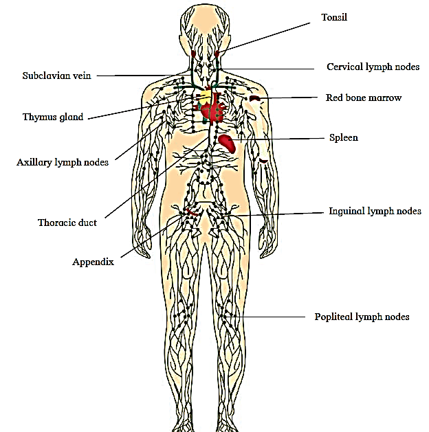
- Lymph, lymphatic capillaries, lymphatic vessels and lymph nodes together constitute lymphatic system.
- Lymph is the tissue fluid that bathes the cells and is collected in lymphatic capillaries.
- Lymph is a fluid connective tissue just like blood but is without RBCs, platelets and some plasma proteins. It contains carbon dioxide and metabolic wastes.
- Lymphatic capillaries are thin walled vessels interwoven with the blood capillaries, present in all the tissue spaces. They are not connected with blood capillaries and are blind at one end. Lymph capillaries are wider than blood capillaries and are lined by endothelium of thin and flat cells.
- Lymphatic vessels are formed by the union of lymphatic capillaries. These are thin walled having numerous valves to prevent backflow. Thoracic or left lymphatic duct and right lymphatic duct are the main lymphatic vessels in the body.
[collapse]
Functions of lymphatic system : Draining off the excess tissue fluid from the extracellular spaces back into the blood.
- Transport of carbon dioxide and metabolic wastes from the tissue fluid. Transport of lymphocytes and antibodies from the lymphatic nodes to the blood.
- Transport of absorbed fats from the intestine to the blood.
- Destruction of invading microorganisms and foreign particles in the lymph nodes.
| Tonsils are small lymphatic nodules in pharyngeal region. Normally there are five tonsils strategically positioned to fight against inhaled and ingested foreign substances. Inflammation of tonsils is called as tonsillitis. It is caused due to viral or bacterial infection. Symptoms include sore throat, fever, swollen lymph nodes, nasal congestion, difficulty in swallowing, headache, etc. Viral tonsillitis cures naturally but bacterial tonsillitis needs antibiotic treatment.
Tonsillectomy is performed in some patients who do not respond to the treatment.
|
Blood and Lymph :
Blood and Lymph :
| Blood |
Lymph |
- Contains blood plasma with proteins and all three types of blood cells namely RBCs, WBCs and blood platelets.
- Red in colour due to presence of RBCs.
- Carries oxygen in the body.
- The flow of blood in blood vessels is fast.
- Lymphocytes are present.
|
Contains blood plasma without blood proteins, RBCs and platelets and contains lymphocytes.
Light yellow in colour and does not contain RBCs.
Does not carry oxygen.
The flow of lymph in lymph capillaries is slow.
Lymphocytes are present, more in number than those present in the blood.
|
[collapse]
<<- Previous Part
Good job 👍
Tq. Kandhar dist. Nanded bahaddarpura