|
Topics to be learn in in Part-1
- The Discovery of DNA
- The Genetic Material is a DNA
- DNA packaging
- DNA replication
- Protein synthesis
Topics to be learn in Part-2 :
- Protein synthesis
- Regulation of gene expression
- Operon concept
- Genomics
- Human Genome Project
- DNA Fingerprinting
|
The Discovery of DNA: Modern information of DNA has evolved from the discovery of nucleic acid to the development of the double-helix model.
Nuclein : (Friedrich Miescher’s nuclein) :
- Nuclein is an acidic substance, having high phosphorus content and it was isolated by Friedrich Miescher in 1869, from the nuclei of pus cells.
- As Nuclein had acidic properties and it was isolated from nucleus, it was called as nucleic acid.
- E Miescher started working with White blood cells (the major component of pus)-
- He used a salt solution to wash the pus off the bandages. He lysed the cells by adding a weak alkaline solution and isolated nucleic acid from nuclei that precipitated out of the solution.
- By the early 1900s, it was known that Miescher’s nuclein was a mixture of proteins and nucleic acids (DNA and RNA).
There are two types of nucleic acids — DNA (deoxyribonucleic acid) and RNA (ribonucleic acid).
The Genetic Material is a DNA : Initially proteins (and not DNA) ' were considered as genetic material because :Proteins are large, complex molecules and store information required to govern cell metabolism. Hence, it was assumed that variations found in species were caused by proteins.
- DNA was considered as a small, simple molecule whose composition varies little among species.
- Variations in the DNA molecules are different than the variation in shape, electrical charge and function shown by proteins.
Various experiments which proved that DNA (and not protein) was the genetic material are as follows :
1-Griffith experiments :
1-Griffith's experiments :
- In 1928, Frederick Griffith, carried out an experiment with two strains of bacterium
- Streptococcus pneumoniae : S-type (Virulent, smooth, pathogenic and encapsulated) and R-type (Non-virulent, rough, non-pathogenic and non-capsulated).
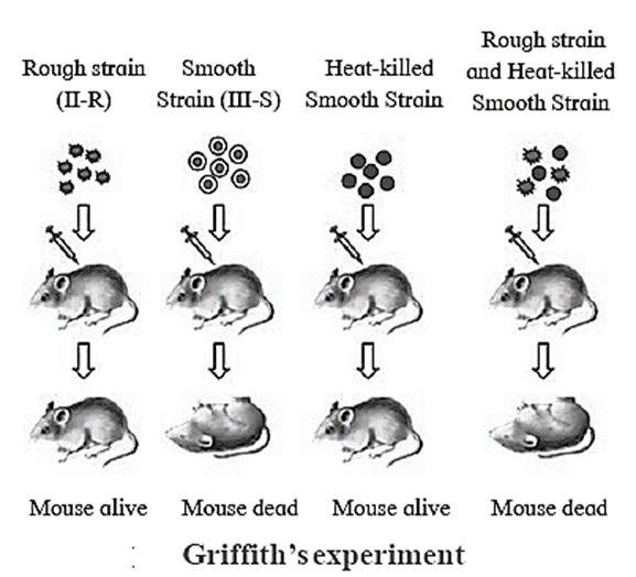
- He observed that on injecting a mixture of heat-killed S bacteria and live R bacteria, the mice died.
- Griffith obtained live S-strain bacteria from the blood of the dead mice.
- Conclusion : Live R-strain bacteria must have picked up something (transforming principle) from the heat-killed S bacterium and got transformed into S-type.
[collapse]
2-Avery, McCarty and MacLeod experiment :
2-Avery, McCarty and MacLeod’s experiment :
- Purified DNA, RNA, proteins, etc. from heat killed cells of S-strain and mixed With R-strain bacteria separately.
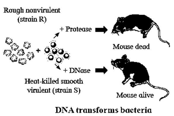
- Only DNA was able to transform avirulent R-strain into virulent S-strain.
- When DNA was digested with DNase, there was no transformation.
- Thus, in 1944, they proved that the DNA is a genetic material (transforming principle), but all biologists were not convinced.
[collapse]
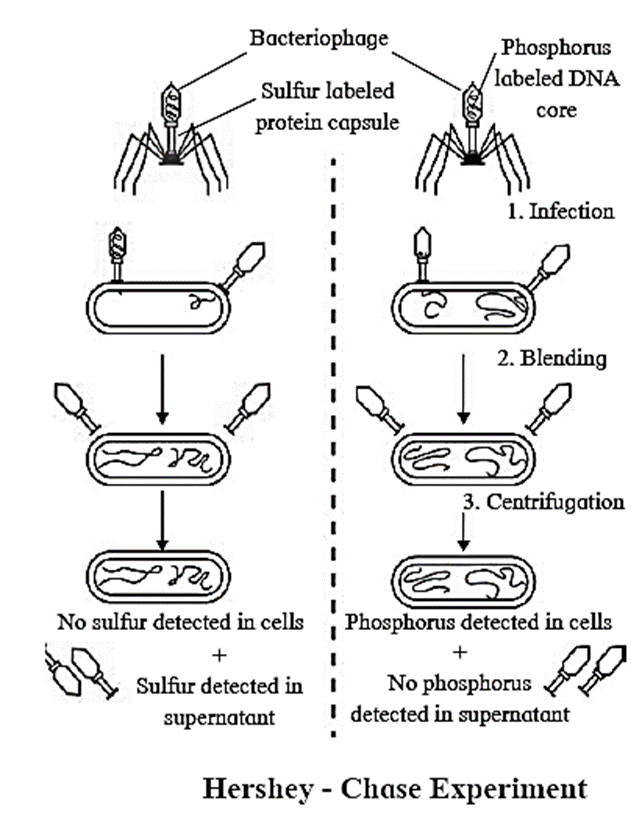
3- Hershey—Chase Experiment :
3- Hershey—Chase Experiment :
- Hershey and Chase worked with bacteriophages.
- Two types of bacteriophages were used in the experiment - type one where DNA was labelled with radioactive phosphorus and type two where protein coat was labelled with radioactive sulphur.
- Steps : infection, blending, centrifugation.
- Experiment proved that DNA is the genetic material which enters bacterial cell and not protein.
[collapse]
DNA packaging : Length of DNA double helix molecule, in a typical mammalian cell is approximately 2.2 meters. Approximate size of a typical nucleus is 10-6 m. This long DNA molecule is accommodated in such a small nucleus. This long DNA molecule is condensed, coiled and super coiled to fit inside such small nucleus.
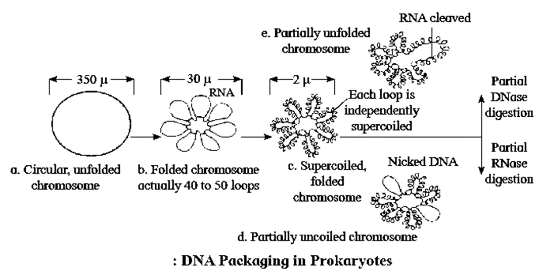
DNA packaging in Prokaryotes :
Packaging in Prokaryotes :
- Size of cell in E. coli size is 2—3 m.
- The nucleoid is small, circular, highly folded, naked DNA (1100, um long in perimeter and contains about 4.6 million base pairs).
- When the negatively charged DNA becomes circular, the size reduces to 850, um in diameter.
- Folding/ looping (40-50 domains (loops) further reduce it to 30, um in diameter.
- RNA connectors assist in loop formation.
- Further coiling and supercoiling of each domain reduces the size to 2 um in diameter.
- This coiling (packaging) is assisted by positively charged HU (Histone like DNA binding proteins) proteins and enzymes like DNA gyrase and DNA topoisomerase-I, which maintain supercoiled state.
[collapse]
DNA packaging in eukaryotic cell :
DNA packaging in eukaryotic cell :
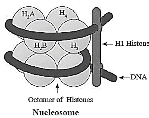
- In eukaryotic cells, DNA (2.2 metres) is condensed, coiled and supercoiled to be packaged efficiently in the nucleus (10-16 m).
- DNA is associated with histone and non-histone proteins.
- Histones are a set of positively charged, basic proteins, rich in basic amino acid residues lysine and arginine.
- Nucleosome consists of nucleosome core (two molecules of each of histone proteins viz. H2A, H2B, H3 and H4 forming histone octamer) and negatively charged DNA (146 bps) that wraps around the histone octamer by 1¾ turns.
- H1 protein binds the DNA thread where it enters and leaves the nucleosome.
- Adjacent nucleosomes are linked with linker DNA (varies in length from 8 to 114 bp. average length of linker DNA is about 54 bp).
- Each nucleosome contains 200 bp of DNA.
- Packaging involves formation of — Beads on string (10 nm diameter), Solenoid fibre (looks like coiled telephone wire. 30 nm diameter/300Å), Chromatin fibre and Chromosome.
- Non-Histone Chromosomal Proteins (NHC) : These are additional sets of proteins contribute to the packaging of chromatin at a higher level.
[collapse]
Heterochromatin and Euchromatin :
Heterochromatin and Euchromatin :
1-Heterochromatin :
- This term was proposed by Heitz.
- Genetically (transcriptionally) almost inactive, darkly stained, condensed parts of chromonema/chromosomes observed in eukaryotic cells, during interphase and early prophase.
- Located near centromere, telomeres are also interrelated.
- Heterochromatin is 2 to 3 times richer in DNA than in the euchromatin.
2-Euchromatin :
- The regions of chromonema which are in non-condensed state, constitute euchromatin.
- Euchromatic regions stain light.
- Euchromatin is genetically (transcriptionally) very much active and fast replicating.
[collapse]
DNA Replication : Replication is the process by which DNA duplicates itself and forms two copies that are identical to it. In eukaryotes, replication of DNA occurs once, in the S-phase of interphase.
Functions of DNA :
- The DNA molecule regulates and controls all the cellular activities.
- DNA replicates and gets distributed equally to the daughter cells when the cell divides.
- Carrier of genetic information.
- Heterocatalytic function : Directs the synthesis of chemical molecules other than itself. E.g. Synthesis of RNA (transcription), synthesis of protein (Translation), etc.
- Autocatalytic function: Directs the synthesis of DNA itself. E.g. Replication.
- A master molecule of a cell that initiates, guides, regulates and controls the process of protein synthesis.
The steps involved in DNA replication :
The steps involved in DNA replication :
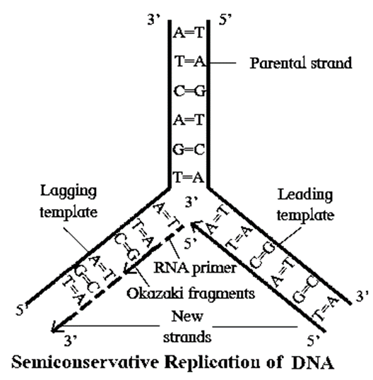
DNA replication is semi-conservative replication. It involves following steps :
1-Activation of Nucleotides :
- Nucleotides (dAMP, dGMP, dCMP and dTMP) present in the nucleoplasm, are activated by ATP in presence of an enzyme phosphorylase.
- This phosphorylation results in the formation of deoxyribonucleotide triphosphates i.e. dATP, dG’l‘R dCTP and dTTP
2-Point of Origin or Initiation point :
- Replication begins at specific point ‘O' Origin and terminates at point ‘T'.
- At the point ‘O’, enzyme endonuclease nicks (breaks the sugar-phosphate backbone or the phosphodiester bond) one of the strands of DNA, temporarily.
3-Unwinding of DNA molecule :
- Enzyme DNA helices breaks weak hydrogen bonds in the vicinity of ‘O’.
- The strands of DNA separate and unwind.
- This unwinding is bidirectional.
- SSBP (Single strand binding proteins) remains attached to both the separated strands and prevent them from recoiling (rejoining).
4-Replicating fork :
- Y-shape replication fork is formed due to unwinding and separation of two strands.
- The unwinding of strands results in strain which is released by super-helix relaxing enzyme.
5-Synthesis of new strands :
- Each separated strand acts as a template for the synthesis of new complementary strand.
- A small RNA primer (synthesized by activity of enzyme RNA primase) get attached to the 3’ end of template strand and attracts complementary nucleotides from surrounding nucleoplasm.
- These nucleotides bind to the complementary nucleotides on the template strand by hydrogen bonds (i.e. A=T or T=A; G=C or C Ξ G, C Ξ G).
- The phosphodiester bonds are formed between nucleotides of new strand to form a polynucleotide strand.
- The enzyme DNA polymerase catalyses synthesis of new complementary strand always in 5’ — 3’ direction.
6-Leading and Lagging strand :
- The template strand with free 3’ is called the leading template.
- The template strand with free 5' end is called the lagging template.
- The replication always starts at C-3 end of template strand and proceeds towards C-5 end.
- New strands are always formed in 5’ -> 3' direction.
- The new strand which develops continuously towards replicating fork is called the leading strand.
- The new strand which develops discontinuously away from the replicating fork is called the lagging strand.
- Maturation of Okazaki fragments : The lagging strand is synthesized in the form of small Okazaki fragments which are joined by enzyme DNA ligase.
- Later RNA primers are removed by the combined action of RNase H, an enzyme, that degrades the RNA strand of RNA-DNA hybrids, and polymerase I.
- Gaps formed are filled by complementary DNA sequence with the help of DNA polymerase-I in prokaryotes and DNA polymerase-a in eukaryotes.
- Finally, DNA gyrase (topoisomerase) enzyme forms double helix to form daughter DNA molecules.
7-Formation of two daughter DNA molecules (semiconservative replication):
- In each daughter DNA molecule, one strand is parental and the other one is newly synthesized.
- Thus, 50% part (i.e. one strand of the helix) is contributed by mother DNA. Hence, it is described as semiconservative replication.
[collapse]
Experimental confirmation of semiconservative replication of DNA :
Experimental confirmation of semiconservative replication of DNA :
Matthew Meselson and Franklin Stahl (1958) used equilibrium — density – gradient — centrifugation technique to experimentally prove semiconservative DNA replication.
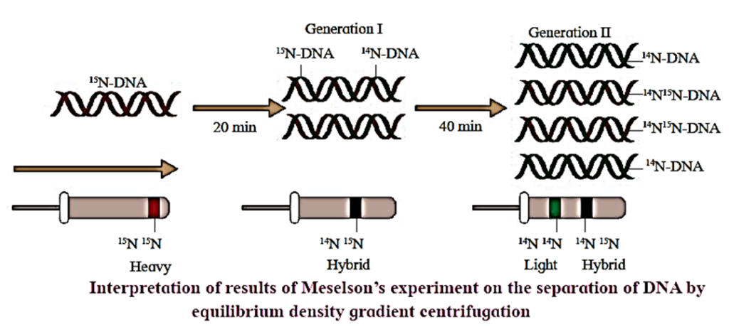
- They cultured bacteria E.coli in the medium containing 14N (light nitrogen). They obtained equilibrium density gradient band by using 6M CsCl2. The position of this band is recorded.
- E. coli cells were then transferred to 15N medium (heavy isotopic nitrogen) and allowed to replicate for several generations.
- At equilibrium point density gradient band was obtained, by using 6M CsCl2. The position of this band is recorded.
- The heavy DNA (15N) molecule can be distinguished from normal DNA by centrifugation in a 6M Cesium chloride (CsCl2) density gradient. At the equilibrium point 15N DNA will form a band. In this both the strands of DNA are labelled with
- 15N.
- Such E. coli cells were then transferred to another medium containing 14N i.e. normal (light) nitrogen. After first generation, the density gradient band for 14N ‘15N was obtained and its position was recorded.
- After second generation, two density gradient bands were obtained — one at 14N ‘15N position and other at 14N position.
- The position of bands after two generations clearly proved that DNA replication is semiconservative.
[collapse]
Next Part ->>
We reply to valid query.