Notes-Part-2
Topics to be Learn : Part-2
|
Muscular tissue :
Characteristics of muscular tissue :
- The cells of the muscular tissue are elongated and are called as muscle fibres.
- The muscle fibres are covered by a membrane called sarcolemma.
- The cytoplasm of the muscle cell is called the sarcoplasm.
- Large number of contractile fibrils called myofibrils are present inthe sarcoplasm.
- Depending on the type of muscle cells, one or many nuclei may be present.
- Myofibrils are made up of the proteins, actin and myosin.
- Muscle fibres contract and decrease in length on stimulation. Hence, muscular tissue is also known as contractile tissue. .
- This tissue is vascular and innervated by nerves.
- Muscle cells contain large number of mitochondria.
Types of Muscular Tissue :
Types of muscles and their locations as follows :
- Skeletal muscles/Striated muscles/Voluntary muscles: They are found attached to bones.
- Smooth / Non-striated muscles/Involuntary muscles: They are found in the walls of visceral organs and blood vessels.
- Cardiac muscles: They are found in the wall of the heart or myocardium.
(i) Skeletal muscles :
There are over 650 named skeletal muscles in the human body. Skeletal muscles are also known as voluntary muscles.
Location: Skeletal muscles are found attached to bones.
Structure:
- They consist of large number of fasciculi which are wrapped by a connective tissue sheath called epimysium or fascia. Each individual fasciculus covered by perimysium.
- Each fasciculus in tum consists of many muscle fibres called myofibers.
- Each muscle fibre is a syncytial fibre that contains several nuclei.

- The sarcoplasm (cytoplasm) is surrounded by the sarcolemma (cell membrane).
- The sarcoplasm contains large number of parallelly arranged myofibrils and hence the nuclei gets shifted to the periphery.
- Each myofibril is made up of repeated functional units called sarcomeres.
- Sarcomere is the functional unit of skeletal muscles
- Each sarcomere has a dark band called anisotropic of ‘A’ band in the centre. ‘A’ bands are made up of the contractile proteins actin and myosin.
- In the centre of the ‘A’ band is the light area called ‘H’ zone or Hensen’s zone.
- In the centre of the Hensen’s zone is the ‘M’ line.
- On either side of the ‘A’ band are light bands called isotropic or ‘I’ bands. These bands contain only actin.
- Adjacent light bands are separated by the ‘Z’ line (Zwischenscheibe line).
- The dark and light bands on neighbouring myofibrils correspond with each other to give the muscles a striated appearance.
Functions: Skeletal muscles show quick and strong voluntary contractions. Skeletal muscles bring about voluntary movements of the body
Different types of skeletal muscles :
Skeletal muscles are divided into two types based on the amount of red pigment (myoglobin).
- Red muscle: It contains very high amount of myoglobin.
- White muscle: It contains very low amount of myoglobin.
Myoglobin : Myoglobin is an iron containing red coloured pigment found only in muscles. It consists of one haeme and one polypeptide chain. It can carry one molecule of oxygen.
Function: Due to presence of myoglobin, the muscles can obtain their oxygen from two sources, myoglobin and haemoglobin.
(ii) Smooth or Non-striated muscles :
Smooth muscles are also known as non-striated, visceral or involuntary muscles.
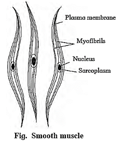
Structure:
- These muscles are present in the form of sheets or layers.
- Each muscle cell is spindle shaped or fusiform.
- The fibres are unbranched and have a single nucleus that is located centrally.
- The sarcoplasm contains myofibrils which are made up of the contractile proteins — actin and myosin.
- Smooth muscles contain less myosin and more actin.
- Striations are absent, hence smooth muscles are also known as non-striated muscles.
- These muscles are innervated by the autonomous newous system.
Location: It is found in walls of visceral organs and blood vessels. Therefore, smooth muscles are also known as visceral muscles. .
Function: Smooth muscles are associated with involuntary movements of the body like peristaltic movement of food through the digestive system.
(iii) Cardiac muscles :
Muscles of the cardiac tissue show characteristics of both striated and non-striated muscle fibres.
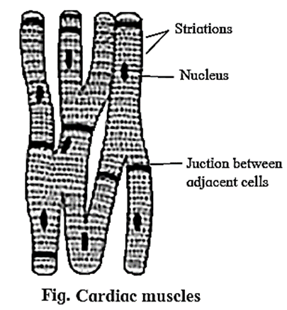
Structure:
- Sarcolemma is not distinct.
- Uninucleate muscle fibres appear to be multinucleate.
- Adjacent muscle fibres join together to give branched appearance.
- Transverse thickenings of the sarcolemma called intercalated discs fonn points of adhesion of muscle fibres.
- These junctions allow cardiac muscles to contract as a unit to aid-quick transfer of stimulus.
Location: They are found in the wall of the heart or myocardium.
Function: Cardiac muscles bring about contraction and relaxation of heart, which helps in circulation of blood throughout the body.
Myogenic heart and Neurogenic heart :
- The mammalian cardiac muscles are modified and are capable of generating an impulse on their own Hence, the mammalian heart known as a myogenic heart.
- In some animals, the cardiac muscles need neural stimulus in order to initiate a contraction. Such a heart is known as a neurogenic heart.
Nervous tissue :
The characteristics of the nervous tissue are as follows:
- The nervous tissue is made up of nerve cells (neurons) and neuroglia.
- Intracellular matrix is absent in the neural tissue.
The neurons are the structural and functional units of the nervous system.
- They are impulse generating and impulse conducting units which bring about quick communication within the body.
- Excitability is the change in action potential of the neuronal membrane on receiving external stimulus.
- Conductivity helps the neurons to carry a Wave of impulse from the dendron to the axon (processes of neuron).
Neuroglial cells : Neuroglial cells are non-nervous supporting cells that fill in the inter-neuronal space and are capable of regeneration and division.
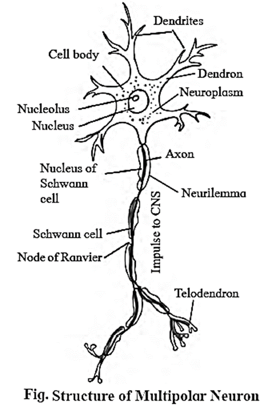
Structure of a neuron :
- A neuron is the structural and functional unit of the nervous tissue. A neuron is made up of cyton or cell body and cytoplasmic extensions or processes.
- The cyton or cell body contains granular cytoplasm called neuroplasm and a centrally placed nucleus.
- The neuroplasm contains mitochondria, Golgi apparatus, RER and Nissl‘s granules.
- Cell body gives out two types of processes namely Dendron and axon.
Dendrons : are short, branched, processes. The fine branches of dendron are called dendrites. They carry impulse towards cyton.
Axon: It is a single, elongated and cylindrical process.
- The axon is bound by the axolemma.
- The protoplasm or axoplasm contains large number of mitochondria and neurofibrils.
- The axon is enclosed in a fatty sheath called the myelin sheath and the outer covering of the myelin sheath is the neurilemma. Both the myelin sheath and the neurilemma are parts of the Schwann cell.
- The myelin sheath is absent at intervals along the axon at the Node of Ranvier.
- The fine branching structure at the end of the axon (terminal arborization) is called telodendron.
Nissl’s Granules :
- Nissls granules are large granular bodies, found in neurons. These granules are made up of rough endoplasmic reticulum (RER) and free ribosomes (site of protein synthesis).
Classification of neurons :
Neurons are classified into three types based on their functions:
- (i) Afferent neuron (Sensory neuron): It carries impulses from sense organ to the central nervous system (CNS). It is found in the dorsal root of the spinal cord.
- (ii) Efferent Neuron (Motor neuron): It carries impulses from CNS to effector organs. It is found in the ventral root of the spinal cord.
- (iii) Interneuron or association neuron: They perform processing, integration of sensory impulses and activate appropriate motor neuron to generate motor impulse. These are located between sensory and motor neurons.
Depending on the presence or absence of myelin sheath neurons are classified into two types:
(i) Myelinated nerve fibre\medullated nerve fibres:
- These nerve fibres have an insulating fatty layer called myelin sheath around the axon. This makes the fibre appear white in colour.
- They have nodes of Ranvier at regular intervals.
- Inter-nodes are present
- Saltatory conduction takes place in myelinated nerve fibres
- These nerve fibres conduct the nerve impulse faster,
- Schwann cell of this nerve fibre secrete myelin sheath.
- Cranial nerves of vertebrates is myelinated.
(ii) Non-myelinated\non-medullated nerve fibres:
- Medullary sheath is absent (Non-myelinated nerve fibre.)
- They do not have nodes of Ranvier,
- Inter-nodes are absent,
- Saltatory conduction is not seen in myelinated nerve fibre.
- These nerve fibres conduct nerve impulse at slow
- These fibres appear grey in colour due to absence Schwann cell of this nerve fibre does not secrete
- Nerves of autonomous nervous system are non-myelinated,
Based on the number of processes given out from cyton, neurons are classified into three types :
(i) Unipolar/Monopolar Neuron :
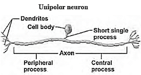
- It has a single process originating from the cyton.
- Both axon and dendron arise from cyton at one point.
- They conduct impulses to central nervous system.
- E.g. Neurons of dorsal root ganglion of spinal nerve.
(ii) Bipolar Neuron :
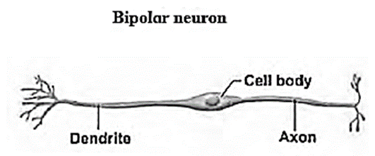
- It has two processes originating from the cyton.
- A single dendron and an axon are given off from opposite poles of the cyton.
- They bring about transmission of special senses like sight, smell, taste, hearing etc.
- E.g. Neurons of retina of eye, olfactory epithelium.
(iii) Multipolar Neuron :
- It is star shaped and gives out more than two processes.
- There is only one axon and remaining are dendrons. Axon initiates from a funnel shaped area called axon-hillock.
- Conduct impulses from receptors to CNS. Integration network between Sensory and motor neuron.
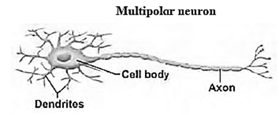
- Almost all neurons in the central nervous system and all motor neurons are multipolar,
Transmission of impulse from one neuron to another :
- A nerve impulse is transmitted from one neuron to another through junctions called synapses.
- A synapse is formed by the membranes of a pre-synaptic neuron and a post-synaptic neuron, which may or may not be separated by a gap called synaptic cleft.
There are two types of synapses, namely, electrical synapses and chemical synapses.
Electrical synapses: The membranes of pre- and post-synaptic neurons are in very close proximity. Thus, electrical current can flow directly from one neuron into the other across these synapses. Impulse transmission across an electrical synapse is faster.
Chemical synapse: The membranes of the pre- and post-synaptic neurons are separated by a fluid-filled space called synaptic cleft.
- Chemicals called neurotransmitters are involved in the transmission of impulses at these synapses.
- The axon terminals contain vesicles filled with these neurotransmitters.
- When an impulse arrives at the axon terminal, it stimulates the movement of the synaptic vesicles towards the membrane where they fuse with the plasma membrane and release their neurotransmitters into the synaptic cleft.
- The released neurotransmitters bind to their specific receptors, present on the post-synaptic membrane. This binding opens ion channels and allows the entry of ions which can generate a new potential in the post-synaptic neuron.
Structure of nerve :
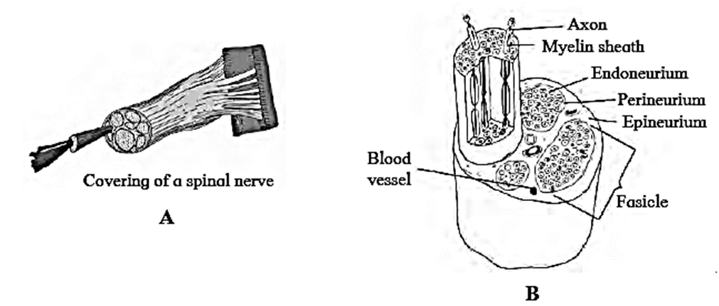
- Each spinal nerve has a large number of axons as well as several layers of connective tissue covers for protection.
- Myelin sheath, a fatty coating, surrounds axons.
- An endoneurium surrounds each axon within a neuron (innermost layer).
- Fascicules are bundles made up of groups of axons along with their endoneurium.
- Each fascicle has perineurium wrapping it (middle layer).
- The epineurium is the nerve's outermost protective layer. There is an epineurium between fascicles.
- The perineurium and epineurium both have numerous blood veins that provide the nerve with nutrition.
We reply to valid query.