Notes-Part-2
Topics to be Learn : Part-2
|
Group of skeleton :
(1) Axial Skeleton : Axial skeleton situated along the vertical axis.
Skull :
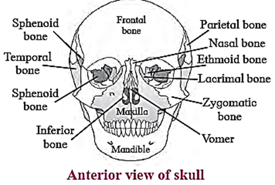
Skull is made up of 22 bones. It is located at the superior end of vertebral column.
The bones of skull are joined by fixed or immovable joints except for jaw. Skull consists of cranium or brain box and facial bones.
Cranium: It is made up of four median bones and two paired bones.
- Frontal bone: It is median bone (unpaired) forming forehead, roof of orbit (eye socket) and the most anterior part of cranium. It is connected to two parietals, sphenoid and ethmoid bone.
- Parietal bones: These paired bones form the roof of cranium and greater portion of sides of the cranium.

- Temporal bones: Theseipaired bones are situated laterally just above the ear on either side. Each temporal bone gives out zygomatic process that joins zygomatic bone to form zygomatic arch. Just at the base of zygomatic process is mandibular fossa, a depression for mandibles (lower jaw bone) that forms the only movable joint of the skull. This bone harbors the ear canal that directs sound waves into the ear. The processes of temporal bones provide points for attachment for various muscles of neck and tongue.
- Occipital bone: It is a single bone present at the back of the head. It forms the posterior part and most of the base of cranium. The inferior part of this bone shows foramen magnum, the opening through which medulla oblongata connects with spinal cord. On the either sides of foramen magnum are two prominent protuberances called occipital condyles. These fit into the corresponding depressions present in 1st vertebra.
- Sphenoid bone: Median bone present at the base of the skull that articulates with all other cranial bones and holds them together. This butterfly shaped bone has a saddle shaped region called sella turcica. In this hypophyseal fossa, the pituitary gland is lodged.
- Ethmoid bone: This median bone is spongy in appearance. It is located anterior to sphenoid and posterior to nasal bones. It contributes to formation of nasal septum and is major supporting structure of nasal cavity.
Facial Bones: Fourteen facial bones give a characteristic shape to the face. The growth of face stops of the age of 16.
Following bones comprise the facial bones:
- Nasals: These are paired bones that form the bridge of nose.
- Maxillae: These form the upper jaw bones. They are paired bones that join with all facial bones except mandible. Upper row of teeth are lodged maxillae.
- Palatines: These are paired bones forming the roof of buccal cavity or floor of the nasal cavity.
- Zygomatic bones: They are commonly called as cheek bones.
- Lacrimal bones: These are the smallest amongst the facial bones.
- These bones form the medial wall of each orbit. They have lacrimal fossa that houses lacrimal sacs. These sacs gather tears and send them to nasal cavity.
- Inferior nasal conchae : They form the part of lateral wall of nasal cavity. They help to swirl and filter air before it passes to lungs.
- Vomer: The median, roughly triangular bone that forms the inferior portion of nasal septum.
- Mandible: This median bone forms the lower jaw. It is the largest and strongest facial bone. It is the only movable bone of skull. It has curved horizontal body and two perpendicular branches i.e. rami. These help in attachment of muscles. It has lower row of teeth lodged in it.
Functions of skull:
- It protects the brain.
- It provides sockets for ear, nasal chamber and eyes.
- Mandible bone of the skull helps in opening and closing of the mouth
Hyoid bone :
- It is a ‘U’ shaped bone that does not articulate with any other bone.
- It is suspended from temporal bone by lingaments and muscles.

- It is located between mandible and larynx.
- It has horizontal body and paired projections called horns.
- It provides site for attachment of some tongue muscles and muscles of neck and pharynx.
| Know This :
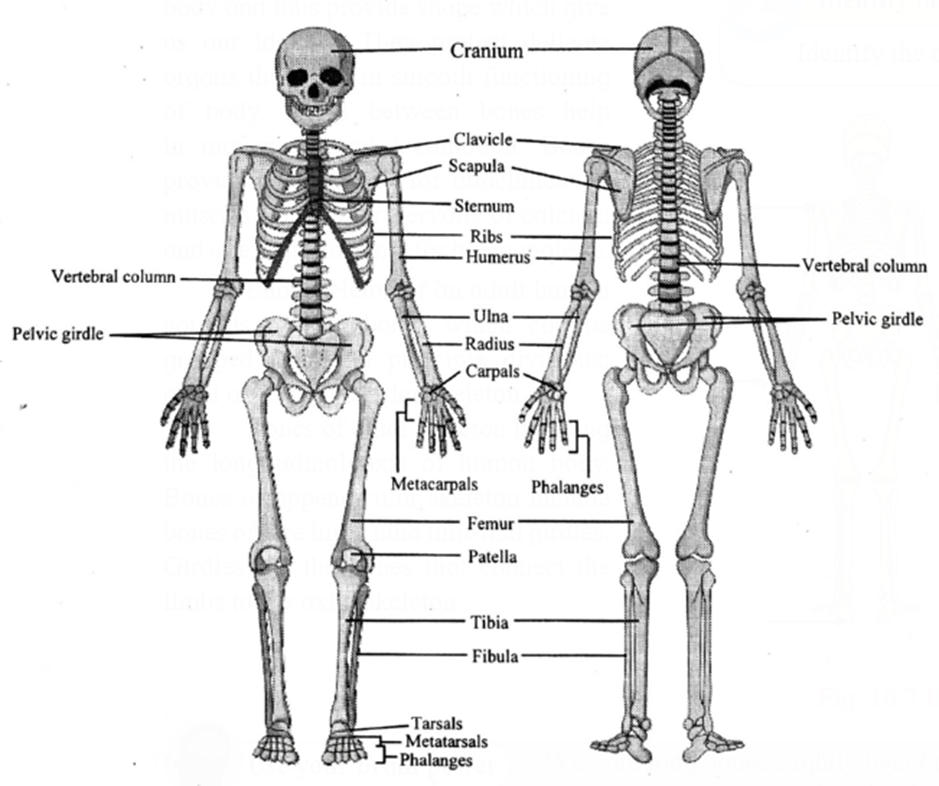
Details of Human Skeleton Axial skeleton :
Subtotal : 80 Appendicular skeleton :
Subtotal : 126 Total : 206 |
Human Skeleton (fig) :
Sutures in skull and their location :
Skull has many sutures (type of immovable joints) present, out of which four prominent ones are
- Coronal suture: Joins frontal bone with parietals.
- Sagittal suture: Joins two parietal bones.
- Lambdiodal suture: Joins two parietal bones with occipital bone.
- Lateral/squamous sutures: Joins parietal and temporal bones on lateral side
Ear ossicles : Three tiny bones namely malleus, incus and stapes, together called ‘ear ossicles’ are present in each middle ear.

Vertebral Column :
- Human vertebral column or backbone is a part of axial skeleton.
- Human backbone or vertebral column is made up of a chain of irregular bones called vertebrae.
- It consists of 33 vertebrae during childhood. In adults, five sacral vertebrae fuse to form one sacrum and four coccygeal vertebrae fuse to form single coccyx, thus total number of bones are 26.
- There are five types of vertebrae, cervical (neck), Thoracic (chest), lumbar (abdominal), sacral (hip region, fused in adults forming sacrum) and coccygeal (fused to form vestigial tail bone called coccyx).

Spine curvatures :
- The spine or vertebral column is curved
- The four curvatures in human spine are cervical, lumbar, thoracic and sacral curvatures.
- The cervical and lumbar curves are secondary and convex whereas the thoracic and sacral curvatures are primary and concave.
- Vertebrae vary in size, shape or processes yet they exhibit similar basic plan.
- Importance of curvature : Curvatures help in balancing in upright position. They absorb shocks while walking and also protect the vertebrae from fracture.
| Know This :
Slipped disc :
|
Typical Vertebra :
Though vertebrae vary in size, shape or processes, they exhibit similar basic plan.
- Each vertebra has prominent central body called centrum.
- The centra of human vertebrae are flat in anterio-posterior aspect. Thus, human vertebrae are amphiplatyan.
- From the either side of the centrum are two thick short processes which unite to form an arch like structure called‘ neural arch, posterior to centrum.
- Neural arch forms vertebral foramen which surrounds the spinal cord.
- Vertebral foramina of all vertebrae form a continuous ‘neural canal‘. Spinal cord along with blood vessels and protective fatty covering passes through neural canal.

- The point where two processes of centrum meet, the neural arch is drawn into a spinous process called neural spine.
- From the base of neural arch, two articulating processes called zygapophyses are given out on either side. The anterior is called superior zygapophyses and posterior called inferior zygapophyses.
- In a stack of vertebrae, inferior zygaphyses of one vertebra articulates with superior zygapophyses of next vertebra. This allows slight movement of vertebrae without allowing them to fall.
- At the junction of zygapophyses, a small opening is fonned on either side of vertebra called inteweitebral foramen that allows passage of spinal nerve.
- From the base of neural arch, lateral processes are given out called transverse processes. Neural arch, neural spine and transverse processes are meant for attachment of muscles.
Atlas vertebrae:

- Atlas is the ring-like, 1st cervical vertebrae. It has anterior, posterior arches and large lateral massesa
- It lacks centrum and spinous process. The superior surfaces of the lateral masses are concave and are known as superior articular facets.
- These facets articulate with the occipital condyles of the occipital bone thereby forming atlanto-occipital joints.
- This articulation permits ‘YES movement’ or nodding movement.
- The inferior surfaces of the lateral masses known as inferior articular facets articulate with axis vertebrae.
Axis vertebra :
- It is the 2nd cervical vertebrae.
- Centrum of this vertebra gives out tooth-like ‘Odontoid Process. Odontoid Process projects superiorly through the anterior portion of the vertebral foramen of the atlas.
- The odontoid process forms a pivot on which the atlas and head rotate. This arrangement allows No movement’ or side to side movement of the head.
- The articulation formed between the anterior arch of atlas, the odontoid process of the axis and between their articular facets is called as atlanto-axial joint.

Importance :
- Vertebral foramen : It houses spinal cord and its meninges
- Odontoid process : This process fits into the anterior portion of vertebral foramen of Atlas vertebra forming pivot joint.
- Inferior articular facet : It articulates with superior articular facet of axis and permits rotatory movement of head.
Typical cervical vertebrae :
- Vertebrae number 3 to 6 are called as typical cervical vertebrae.
- They show short centrum and bifid spinous process.
- The transverse processes of these vertebrae are reduced; each having large vertebrarterial canal at its base for the passage of vertebral artery.
7th cervical vertebra (Vertebra prominens): It is the largest cervical vertebra where the neural spine is straight.
Thoracic vertebra :
- These are twelve in number and found in chest region.
- Centrum of thoracic vertebrae is heart shaped and all processes are well developed. Except for vertebrae number 11th and 12th transeverse process of other thoracic vertebrae show facets for attachment with ribs.
- Thoracic vertebrae can be identified on the basis of centrum as the centrum of the thoracic vertebrae is heart shaped.

Lumbar vertebra :
- There are five lumbar vertebrae.
- These are well developed vertebrae that exhibit all characters of a typical vertebra.
- The centrum of the lumbar vertebrae is kidney shaped.

Sacrum:
- It is a triangular bone formed by the fusion of five sacral vertebrae.
- It is located in pelvic cavity between two hip bones.
- The anterior end of sacrum is broad and posterior end is narrow.
- It consists of vertebral foramina formed by the fusion of vertebrae.
- The reduced neural spines can be observed projecting from dorsal aspect of sacrum.
- Function: It gives strength to pelvic girdle.
Coccyx:
- It is a triangular bone which is formed by fusion of four coccygeal vertebrae.
- It is reduced and does not show vertebral foramina and spinous processes.
- The transverse processes of coccygeal vertebrae are reduced.

Thoracic cage :
Thoracic cage consists of 12 pairs of ribs, breast bone or sternum.
Sternum:

- It is a flat, narrow bone about 15 cm long.
- It is positioned medially in the anterior thoracic wall (chest region).
- It is divided into three sections: the manubrium, the body, and the xiphoid processes.
- Manubrium has two notches on the anteriolateral side for attachment to each side's clavicle. It also depicts two notches on each lateral side for the attachment of the first two pairs of ribs.
- The sternum body is a flat bone with five notches on the lateral aspect for direct or indirect rib attachment.
- The xiphoid process is the lowermost part of the sternum that is initially cartilaginous and ossifies in adults. It allows for the attachment of the diaphragm and abdominal muscles.
- Ribs are attached to sternum by means of cartilaginous extensions called costal cartilages.
Ribs:

- A rib is a bone with a 'C' shape. It is attached to the corresponding thoracic vertebrae on the dorsal side.
- There are twelve pairs of ribs attached to twelve thoracic vertebrae. The posterior ends of the ribs have two protuberances for attachment to the vertebrae: the head and tubercle.
- The head of the rib attaches to a facet formed by demifacets of adjacent thoracic vertebrae at the base of transverse processes.
- The tips of these vertebrae's transverse processes also have facets for rib attachment where tubercles of ribs are attached.
- Ribs provide a location for intercostal muscles to attach. The space between the ribs is called as intercostal space
- On ventral side, the ribs may or may not attach the sternum. Depending on their attachment, the ribs are classified into three types:
- True ribs: First seven pairs of ribs are attached directly to the sternum by means of their costal cartilages.
- False ribs: Costal cartilages of rib numbers 8, 9 and l0 are attached to rib number 7 on either side and not directly to the sternum. Hence, these are called false ribs.
- Floating ribs: Last two pairs of ribs have no ventral connection. Hence, they are called floating ribs.
| Know This :
Approximately 8 % of humans have an extra pair of ribs attached to the lumbar vertebra. Such a rib is found in some types of gorillas. Hence 13th pair of ribs is called gorilla rib. |
(2) Appendicular skeleton :
Appendicular skeleton consists of bones of limbs and girdles.
Pectoral girdle :
- The pectoral girdle, also known as the shoulder girdle, connects the forelimb skeleton to the axial skeleton.
- Each pectoral girdle is made up of a shoulder blade or scapula and a collar bone or clavicle.

Clavicle :
- It is ‘s’ shaped slender bone.
- One end of the clavicle is connected to the acromion process of the scapula.
- The other rounded end, known as the sternal end, connects to the manubrium of the sternum.
- This connects the upper arm and axial skeletons.
Scapula:
- It is a large, flat, triangular bone that occupies the posterior chest wall from the second to the seventh ribs and is attached to the axial skeleton by muscles and tendons.
- At its lateral angle, the scapula bears a concave socket called the glenoid cavity, into which the head of the humerus (the upper arm bone) fits. It is located in shoulder and hips.
- A beak-like coracoid process projects from the scapula, and an acromion process arises from the scapula. They can be felt as a high point of the shoulder. Both processes are intended for muscle attachment.
Bones of forelimb :
It is made up of 30 bones, including the humerus, radius, and ulna (which form the forearm bones), wrist bones (carpals), palm bones (metacarpals), and digitsphalanges.
(i) Humerus:

- This is the bone of upper arm.
- It has a hemispherical head at its proximal end. On either side of head of humerus are present a pair of projections termed greater and lesser tubercles.
- Bicipital groove is a deep groove present between the tubercles where a tendon of biceps muscle is attached.
- The shaft of humerus shows deltoid tuberosity. Distal end of humerus shows pulley like part called trochlea that articulates with ulna.
(ii) Radius and Ulna:

- Radius is located on the thumb side of the forearm, laterally.
- The radius's proximal end has a disc-like head that articulates with the humerus bone.
- The radius shaft widens distally to form the styloid process.
- The ulna is located on the forearm's little finger side, medially.
- The Olecranon process, located at the proximal end of the ulna, forms the elbow joint with the humerus bone. On the lateral side, near the upper end of the ulna, there is a radial notch into which the side of the radius head is fixed.
- Radius and ulna articulate with each other at upper and lower extremities by superior and inferior radio-ulnar joints. In between the shafl of two bones, interosseous membrane is present.
(iii) Carpals: These are bones of wrist, arranged in two rows of four each.
(iv) Metacarpals: Five elongated metacarpals form -the bones of palm. Their proximal ends join with carpals and distal ends form knuckles.
(v) Phalanges: Phalanges form the bones of fingers and thumb. There are 14 phalanges in each hand (Four fingers have three phalanges each and thumb has two).

Pelvic girdle : Pelvic girdle also known as hip girdle connects hind limb skeleton with axial skeleton.

- It is composed of two hip bones known as coxal bones. These coxal bones join the sacrum posteriorly.
- The coxal bone is a large irregularly shaped bone composed of three parts: the ilium, the ischium, and the pubis.
- At the point of fusion of the ilium, ischium, and pubis, there is a cavity called the acetabulum, which forms a ball and socket joint with the thigh bone.
- The pubic symphysis is a cartilaginous joint that connects the two pubis bones medially. The pubis and ischium together form a bone ring that encloses the obturator foramen.
Bones of lower limb:
The bones of lower limb are femur, patella, tibia and fibula, tarsals, metatarsals and phalanges
(i) Femur: The thigh bone is the longest bone in the body. The head is joined to shaft at an angle by a short neck. It forms ball and socket joint with acetabulum cavity of coxal bone. The lower one third region of shaft is triangular flattened area called popliteal surface. Distal end has two condyles that articulate with tibia and fibula.
(ii) Patella: It is also called as knee cap. It is a sesamoid bone (bone embedded in tendon). It is a flat rounded bone with a pointed lower end.

(iii) Tibia and fibula: These are the two long bones of shank or lower leg. The two bones are connected to each other at the extremities. In between the two bones interosseous membrane is present.
- Tibia: It is much thicker and stronger than fibula. Its broad and expanded upper end articulates with femur and the lower end articulates with talus (tarsal bone).
- Fibula: It is a long slender bone on lateral side of tibia.

(iv) Tarsals: These are the bones of ankle. Seven tarsals are arranged in three rows, two proximal, one intermediate and four distal.
(v) Metatarsals: Five metatarsal bones support the foot. Proximally they attach with distal row of tarsals and distally the metatarsals articulate with phalanges. .
(vi) Phalanges: These are the bones of the toes. Except the big toe which has two phalanges, the other four toes have three phalanges each.
Types of joints :
Joint : A point where two or more bones get articulated is called joint or articulation or athrosis. The study of joints is called arthrology.
They are classified based on degree of flexibility or movement they permit into lastly synovial or freely movable or diaithroses type of joints.
Synarthroses / fibrous joints / movable joints:
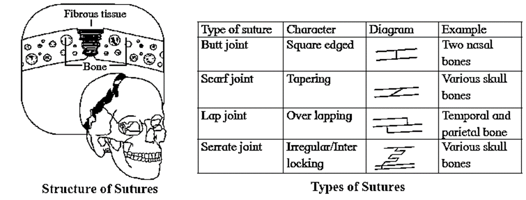
In this joint, the articulating bones are held together by means of fibrous cormective tissue. Bones do not exhibit movement. Hence, it is immovable or fixed type of joint. Synarthroses are further classified into sutures, syndesmoses and gomphoses.
- Sutures: It is composed of thin layer of a dense fibrous connective tissue. Sutures are places of growth. They remain open till growth is complete. On completion of growth, they tend to ossify. Sutures may permit some moulding during childhood. Sutures are further classified into butt joint, scarf joint, lap joint and serrate joint.
- Syndesmoses: It is present where there is greater distance between articulating bones. At such locations, fibrous connective tissue is arranged as a sheet or bundle. e.g. Distal tibiofibular joint, interosseous membrane between tibia and fibula and that between radius and ulna.
- Gomphoses: In this type of joint, a cone shaped bone fits into a socket provided by other bone. e.g. Tooth and jaw bones.

Cartilaginous / slightly movable joints / amphiarthroses:
These joints are neither fixed nor freely movable. Articulating bones are held together by hyaline or fibrocartilages. They are further classified as
- Synchondroses: The two bones are held together by hyaline cartilage. They are meant for growth. On completion of growth, the joint gets ossified. e.g. Epiphyseal plate found between epiphysis and diaphysis of a long bone, Rib - Sternum junction.

- Symphysis: In this type of joint, broad flat disc of fibrocartilage connects two bones. It occurs in mid-line of the body. e.g. Intervertebral discs, manubrium and sternum, pubic symphysis.

Synovial joints / freely movable joints / diarthroses:

- It is characterized by presence of a space called synovial cavity between articulating bones that renders free movement at the joint.
- The articulating surfaces of bones at a synovial joint are covered by a layer of hyaline cartilage. It reduces friction during movement and helps to absorb shock.
- Synovial cavity is lined by synovial membrane that forms synovial capsule. Synovial membrane secretes synovial fluid.
- Synovial fluid is a clear, viscous, straw coloured fluid similar to lymph. It is viscous due to hyaluronic acid. The synovial fluid also contains nutrients, mucous ‘and phagocytic cells to remove microbes.
- Synovial fluid lubricates the joint, absorbs shocks, nourishes the hyaline cartilage and removes waste materials from hyaline cartilage cells (as cartilage is avascular). Phagocytic cells destroy microbes and cellular debris formed by wear and tear of the joint.
- If the joint is immobile for a while, the synovial fluid becomes viscous and as joint movement starts, it becomes less viscous.
- The joint is provided with capsular ligament and numerous accessory ligaments. The fibrous capsule is attached to periosteum of articulating bones. The ligament helps in avoiding dislocation of joint.
The types of synovial joints are on follows:
- Pivot joint: In this type of joint, the rounded or pointed surface of one bone articulates with a ring formed partly by another bone and partly by the ligament. Rotation only around its own longitudinal axis is possible. e.g. In joint between atlas and axis vertebrae, head tums side ways to form ‘NO’ joint.
- Ball and socket joint: The ball like surface of one bone fits into cup like depression of another bone forming a movable joint. Multi-axial movements are possible. This type of joint allows movements along all three axes and in all directions. e.g. Shoulder and hip joint.

- Hinge joint: In a hinge joint, convex surface of one bone fits into concave surface of another bone. In most hinge joints one bone remains stationary and other moves. The angular opening and closing motion (like hinge) is possible. In this joint only mono-axial movement takes place like flexion and extension. e.g. Elbow and knee joint.
- Condyloid joint: It is an ellipsoid joint. The convex oval shaped projection of one bone fits into oval shaped depression in another bone. It is a biaxial joint because it permits movement along two axes viz. flexion, extension, abduction, adduction and circumduction is possible. e.g. Metacarpophalangeal joint.

- Gliding joint: It is a planar joint, where the articulating surfaces of bones are flat or slightly curved These joints are non-axial because the motion they allow does not occur along an axis or a plane. e.g Intercarpal and intertarsal joints.
- Saddle joint: This joint is a characteristic of Homo sapiens. Here the articular surface of one bone is saddle-shaped and that of other bone fits into saddle (each bone forming this joint have both concave and convex areas). It is a modified condyloid joint in which movement is somewhat more free. It is a biaxial joint that allows flexion, extension, abduction, adduction and circumduction. e.g. Carpometacarpellar joint between carpal (trapezium) and metacarpal of thumb.

| Know This :
Tennis elbow : • Tennis elbow is caused by inflammation of the tendon that connects the forearm muscles to the upper arm bone (humerus). • It causes excruciating pain in the elbow. It is caused by extensive repetitive hand movement. This causes tendon damage and increases elbow joint tenderness. • This disorder affects not only athletes, but also regular people whose jobs require extensive hand movement, such as carpenters, painters, plumbers, and so on. |
Disorders related to muscles :
Muscular dystrophy:
- It is a genetic disorder that causes gradual wasting of various muscle groups. Typically, the voluntary skeletal muscles are weakened, whereas internal muscles such as the diaphragm are not affected in patients suffering from this disorder.
- Duchenne muscular dystrophy, which affects the lower limbs, is more common in boys.
- Limb girdle muscular dystrophy affects the muscles of the shoulders or hips and typically begins in adults between the ages of 20 and 30.
- This disease has no known cure.
Myasthenia gravis:
- It is a weakness of skeletal muscles.
- It is caused by an abnormality at the neuromuscular junction that partially blocks muscle contraction.
- It is an autoimmune disorder caused by excessive production of certain antibodies in the blood stream. These antibodies bind to acetylcholine receptors of neuromuscular junction. Thus, the transmission of nerve impulses to the muscle fibres is blocked. This causes progressive and extensive muscle weakness.
- It may affect the eye and eyelid movements, facial expression and swallowing.
- The degree of muscle weakness varies fonn local to general.
- Symptoms : Ptosis (drooping or falling of upper eye lid), diplopia or double vision, difficulty in swallowing, chewing and speech.
Disorders related to bones :
Arthritis: It is an inflammation of joints. It is a painful disorder of bones, ligaments, tendons, etc. In this disorder, joints become swollen, stiff and painful. It can lead to disability.
Arthritis is of three types:
- Osteoarthritis: In this disorder, the joint cartilage is degenerated. It is caused by various factors like aging, obesity, muscle weakness, etc. This is the most common type of arthritis that affects hands, knees and spine.
- Gouty arthritis (Gout): In this disorder, joint pain occurs due to the deposition of uric acid in joints. If uric acid is produced in excess or is not excreted, it accumulates in joints as sodium urate which degenerates the cartilage, causing inflammation and pain. It generally affects the joints of feet.
- Rheumotoid arthritis: It is an autoimmune disorder where body’s immune system attacks its own tissues. In rheumotoid arthritis, synovial membrane swells up and starts secreting extra synovial fluid. This fluid exerts pressure on joint and makes it painful. Membrane may develop abnonnal granulation tissue called pannus. Pannus may erode cartilage. Fibrous tissue gets ossified and may lead to stiffness in joints.
Osteoporosis:
- In this disorder, bones become porous and hence brittle. It is primarily age related disease and is more common in women than men.
- As age advances, bone resorption outpaces bone formation. Hence, the bones lose mass and become brittle. More calcium is lost in urine, sweat, etc., than it is gained through diet. Thus, prevention of disease is better than treatment by consuming adequate amount of calcium and exercise at young age.
- Osteoporosis may be caused due to decreasing estrogen secretion after menopause, deficiency of vitamin D, low calcium diet, decreased secretion of sex hormones and thyrocalcitonin.
We reply to valid query.