Notes-Part-1
|
Topics to be Learn : Part-1
Topics to be Learn : Part-2
|
Introduction :
Tissue : A group of cells with the same embryonic origin, structure, and function is called a tissue.
Organs : Organs are large functional units made up of a variety of tissues that come together in an organised way. E.g. Kidneys
Organ-system: Number of organs combine together to form an organ-system. e.g. Respiratory system
Cells :
Number of cells in human body : There are about 100 trillion of 200 different types of cells in the human body.
In a multicellular organism, cells are broadly classified into two types: (i) Somatic cells (ii) Germ cells
- Somatic cells: All body cells except the sperm and the ova are called as somatic cells.
- Germ cells: The sperm and the ova are known as germ cells. They are related to reproductive system
![]()
Histology
Histology : The study of the structure coelom, etc. and arrangement of tissue is called histology.
Types of animal tissues : Animals have four different types of tissues: epithelial, connective, muscular, and nervous.
| Know This :
Marie Francois Xavier Bichat (1771-1802), French anatomist and pathologist discovered tissue. He was known as ‘Father of Histology’. |
Epithelial tissue : (epi : above, thelium : layer of cells)
Characteristics of epithelial tissue :
The characteristics of epithelial tissues are as follows:
Structure:
- This tissue's cells are tightly packed together and have minimal intercellular matrix.
- A non-cellular basement membrane supports the cells.
- The form of the epithelial cells is polygonal, cuboidal, or columnar.
- A single nucleus is present at the centre or at the base of the cell.
- The tissue is avascular and has a good regeneration capacity.
Function:
- Protection is the epithelial tissue's primary function.
- Additionally, it helps in filtration, secretion, absorption, and transportation.
Types of epithelial tissues :
The different types of epithelial tissues are as follows:
Simple epithelium: Epithelial tissue madeup of single layer of cells is known as simple epithelium. Simple epithelium is further classified into:
- Squamous Epithelium
- Cuboidal Epithelium
- Columnar Epithelium
- Ciliated Epithelium
- Glandular Epithelium
- Sensory epithelial tissue
- Genninal epithelial tissue
Compound epithelium: Epithelium composed of several layers is called compound epithelium. Compound epithelium is further classified into:
- Stratified epithelium
- Transitional epithelium
Basement membrane : Basement membrane is a non - cellular membrane on which the lowermost layer of the epithelium lies. Epithelial tissue rests on a basement membrane which acts as a scaffolding on which epithelium can grow and regenerate after injuries.
(A) Simple epithelium :
(i) Squamous epithelial tissue :

Location: It is present in blood vessels, alveoli, coelom, etc.
Structure:
- The squamous epithelium is composed of single layer of cells.
- The cells are polygonal in shape, thin and flat, with serrated margin.
- They have centrally placed spherical or oval nucleus.
- They appear like flat tiles when viewed from above, thus, are also called as pavement epithelium
Functions: Protection, absorption, transport, filtration and secretion.
(ii) Cuboidal epithelium :
Location: It is present in the lining of pancreatic ducts, salivary duct, proximal and distal convoluted tubules of nephron, etc.
Structure:
- The cells are cuboidal in shape.
- They have a centrally placed, spherical nucleus.
Functions: Absorption and secretion.

(iii) Columnar Epithelium:
Location: It is found in inner lining of intestine, gall bladder, gastric glands, intestinal glands, etc.
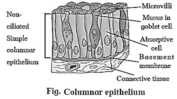
Structure:
- The cells are tall, pillar-like. The inner ends of the cells are narrow while free ends are broad and flat.
- Nucleus is oval or elliptical in the lower half of the cell.
- Free surface shows large number of microvilli.
Function: Secretion and absorption.
(iv) Ciliated epithelium :
Location: It is found in inner lining of buccal cavity of frog, nasal cavity, trachea, oviduct of vertebrates, etc.
Structure:
- Cells of this tissue are cuboidal or columnar.
- Free ends of cells are broad while narrow ends rest on a basement membrane.
- The free ends of the cell show hair-like cilia.
- The nucleus is oval and placed at basal end of the cell.
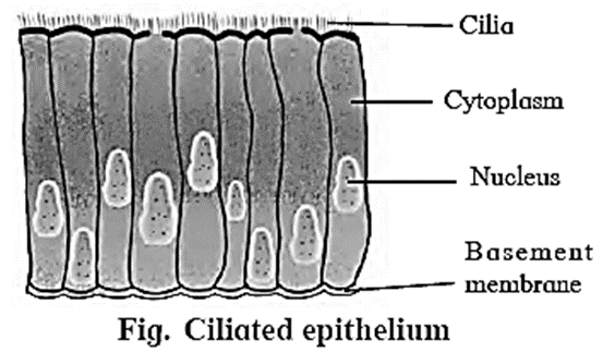
Function: To create a movement of materials that come in contact with the epithelium, in a specific direction. This aids in functions like prevention of entry of foreign particles in the trachea, pushing of the ovum through the oviduct, etc.
(v) Glandular epithelium :
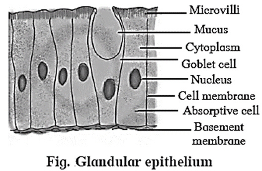
Structure:
- The cells of the glandular epithelium can be columnar, cuboidal or pyramidal in shape.
- The nucleus of these cells is large and situated towards the base.
- Secretory granules are present in the cell cytoplasm.
- The glands may be either unicellular (goblet cells of intestine) or multicellular (salivary gland), depending on the number of cells.
Types: Depending on the mode of secretion, multicellular glands can be further classified as duct bearing glands (exocrine glands) ad ductless glands (endocrine glands).
- Exocrine glands: These glands pour their secretions at a specific site. e.g. salivary gland, sweat gland, etc.
- Endocrine glands : These glands release their secretions directly into the blood stream. e.g. thyroid gland, pituitary gland, etc.
Function: Glandular epithelium secrete mucus to trap the dust particles, lubricate the inner surface of respiratory and digestive tracts, secrete enzymes and hormones, etc.
(vi) Sensory epithelial tissue :
Location: It is found in the nose (Olfactory), ear (Auditory hair cells) and eye (photoreceptors).

Structure :
- Sensory epithelial tissues are composed of a modified form of columnar cells and elongated neurosensoiy cells.
- Sensory hairs are present at the free end of these cell.
Function: It perceives external as well as internal stimuli.
(vii) Germinal epithelium :
Function : Cells of this epithelium divide meiotically to produce haploid gamets. Ex. : Lining of seminiferous tubules, inner lining of ovary.
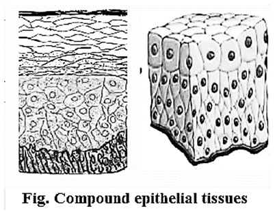
(B) Compound epithelium :
- Compound epithelium consists of many layers of cells.
- Only the lowermost layer of this tissue is based on the basement membrane.
Types of compound epithelium include:
(i) Stratified epithelium:
- Nucleus is present in stratum germinativum (basal layer).
- Cells at free surface become flat and lack nucleus called stratum corneum.
Function: Protection. e.g. Epidermis of skin, oesophagus, cornea, vagina, rectum.
(ii) Transitional epithelium:
- Structure of transitional epithelium is same like stratified epithelium.
- The cells can undergo a change in their shape and structure depending on degree of stretch.
- As the tissue stretches, the transitional cells start changing shape from round and globular to thin and flat.
Function: Distension of organ e. g. Urinary bladder.
Distinguish between simple epithelium and compound epithelium :
| Simple epithelium | Compound epithelium |
| It is made up of single layer of cells | It is made up of two or more layer of cells |
| Single layer of cells that rest on the basement membrane. | Only lowermost layer rests on the basement membrane. |
| It is useful in diffusion, osmosis, filtration, secretion and absorption. | Generally protective in function. It has limited role in absorption. |
| It is generally present in the outer and inner lining of organs, blood vessels etc. | It is present in the epidermis of skin, oesophagus, cornea, vagina, rectum, urinary bladder, etc. |
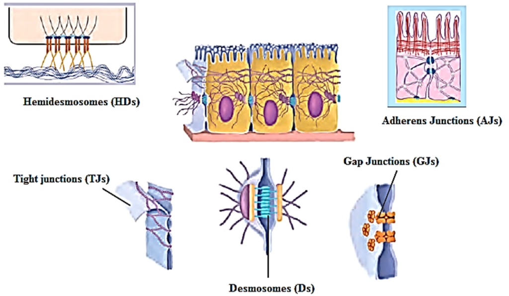
Cell junctions : The epithelial cells are connected to each other laterally as well as to the basement membrane by junctional complexes called cell junctions.
The different types of cell junctions are as follows:
- Gap Junctions (GJs): These are intercellular connections that allow the passage of ions and small molecules between cells as well as exchange of chemical messages between cells.
- Adherens Junctions (AJs): They are involved in various signalling pathways and transcriptional regulations.
- Desmosomes (Ds): They provide mechanical strength to epithelial tissue, cardiac muscles and meninges.
- Hemidesmosomes (HDs): They allow the cells to strongly adhere to the underlying basement membrane. These junctions help maintain tissue homeostasis by signalling.
- Tight junctions (TJs): These junctions maintain cell polarity, prevent lateral diffusion of proteins and ions.
Connective tissue :
Connective tissue is the most widely spread tissue in the body which binds, supports and provides strength to other body tissues and organs.
Characteristics:
- It consists of a variety of cells and fibres which are embedded in the abundant intercellular substance called matrix.
- Fibroblasts are the cells of connective tissue which form fibres
- It is a highly vascular tissue, except cartilage.
The connective tissue is classified on the basis of matrix present, into three types namely (i) connective tissue proper, (ii) supporting connective tissue (iii) fluid connective tissue.
(i) Connective tissue proper is further classified as
- Loose connective tissue (e.g. areolar connective tissue and adipose tissue)
- Dense connective tissue (e.g. ligament and tendon).
(ii) Supporting connective tissue also called skeletal tissue includes cartilage and bone.
(iii) Fluid connective tissue includes blood and lymph.
Functions: Connective tissue protects the vital organs of the body. It acts as packing material and also helps in healing process.
(A) Connective Tissue Proper :
(1) Loose connective tissue : Matrix of loose connective tissue is semisolid, jelly like, viscous matter made up of gelatin.
(i) Areolar connective tissue :

Location : Areolar tissue is a loose connective tissue found under the skin, between muscles, bones, around organs, blood vessels and peritoneum. It is composed of fibres and cells.
The matrix of areolar tissues contains two types of fibres i.e. white fibres and yellow fibres.
- White fibres: They are made up of collagen and give tensile strength to the tissue.
- Yellow fibres: They are made up of elastin and are elastic in nature.
The four different types of cells present in this tissue are as follows:
- Fibroblast: Large fiat cells having branching processes. They produce fibres as well as polysaccharides that form the ground substance or matrix of the tissue.
- Mast cells: Oval cells that secrete heparin and histamine.
- Macrophages: Amoeboid, phagocytic cells.
- (Fat cells) Adipocytes: Cells that store fat. These cells have eccentric nucleus.
Function of areolar tissue :
Areolar tissue acts as packing material, helps in healing process and connects different organs or layers of tissues.
(ii) Adipose tissue (adipo = fat):
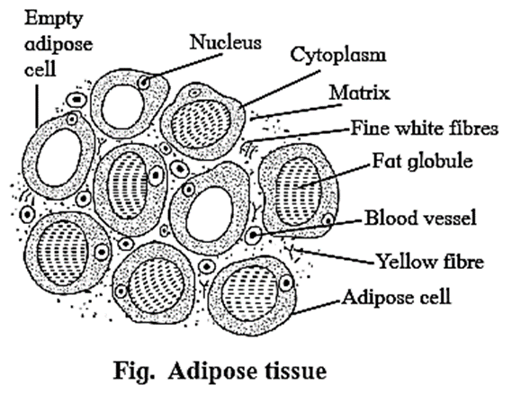
Location: It is found in association with areolar connective tissue. Adipose tissue is present beneath the skin, around the kidneys and between internal organs.
Structure:
- It contains large number of adipocytes.
- The cells are rounded or polygonal.
- Due to presence of fats stored in the form of droplets in adipocytes, the nucleus is shifted towards the periphery.
- Matrix is less and fibres and blood vessels are few in number.
The adipose tissue is of two types:
(i) White adipose tissue:
- It is opaque due to the presence of large number of adipocytes.
- It is commonly present in adults.
(ii) Brown adipose tissue:
- It is reddish brown in colour due to the presence of large number of blood vessels.
Functions:
- Adipose tissue is a good insulator, acts as a shock absorber and a good source of energy because it stores fat.
- The tissue is found in the sole and palm region as well as around organs like kidneys.
- The adipose tissue acts as good insulator and helps retain heat in the body. This helps in survival of animals in the colder regions.
(2) Dense Connective Tissue :
Fibres and fibroblasts are compactly arranged in the dense connective tissue.
There are two types of dense connective tissue :
- Dense regular connective tissue: Collagen fibres are arranged in a parallel manner. e.g. Tendons and ligaments
- Dense irregular connective tissue: Fibres and fibroblasts are not arranged in an orderly manner. E.g. Dermis of skin.
Tendons :
- Tendons are a type of dense regular connective tissue.
- Tendons connect skeletal muscles to bones.
- They contain bundles of white fibres which give tensile strength to the tissue e.g. Achilles tendon, Hamstring tendon
Achilles tendons (Calcaneal tendons) connect the calf muscles to the heel bone.
- When the calf muscles flex, the Achilles tendon pulls on the heel. This movement allows us to stand on our toes.
- Generally, a pain at the back of ankle or lower calf may signal a problem with an Achilles Tendon.
- Athletes who participate in track and field may face Achilles tendon injury.
- The Achilles tendon is the largest and strongest tendon in the body.
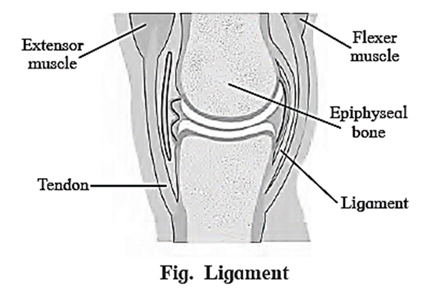
Ligaments :
- Ligaments are a type of dense regular connective tissue that are made up of elastic or yellow fibres arranged in regular pattern. These fibres make the ligaments elastic.
- Location: Ligaments are present at joints.
- Function: Ligaments prevent dislocation of bones.
(C) Supporting connective tissue :
Supporting connective tissue is a type of connective tissue which is characterised by the presence of hard matrix.
It is classified into two types i.e., cartilage and bone.
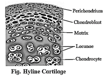
(i) Cartilage : Cartilage is a type of supporting connective tissue. It is a pliable yet tough tissue.
Structure:
- Abundant matrix is delimited by a sheath of collagenous fibres called perichondrium.
- The matrix is called chondrin.
- Below the perichondrium, immature cartilage forming cells called chondroblasts are present.
- Chondroblasts mature and get convened into chondrocytes.
- Chondrocytes are scattered in the matrix and are enclosed in the lacunae
- Each lacuna contains 2 to 8 chondrocytes.
- It forms the endoskeleton of cartilaginous fishes like shark.
- It is widely distributed in vertebrate animals
Based upon the type of matrix, there are four types of cartilage as explained below.
1-Hyaline cartilage:
- The hyaline cartilage is elastic and compressible in nature.
- Perichondrium is present in this cartilage.
- Its matrix is bluish white and gel like.
- Very fine collage fibres and chondrocytes are present in this cartilage.
- Location: It is found at the end of long bones, epiglottis, trachea, ribs, larynx and hyoid.
- Function: It acts as a good shock absorber as well as provides flexibility. It reduces friction.
2-Elastic cartilage:
- The perichondrium is present in elastic cartilage.
- The matrix contains elastic fibres and chondrocytes are few in numbers.
- Function: It gives support and maintains shape of the body part.
- Location: It is found in the ear lobe, tip of the nose, etc.

3-Fibrocartilage:
- The fibrocartilage is the most rigid cartilage.
- Perichondrium is absent in the fibrocartilage.
- The matrix contains bundles of collagen fibres and few chondrocytes that are scattered in the fibres.
- Function: It maintains position of vertebrae.
- Location: Intervertebral discs are made up of fibrocartilage. It is also found at the pubic symphysis.

4-Calcified cartilage:
This type of cartilage becomes rigid due to deposition of salts in the matrix, reducing the flexibility of joints in old age. E.g- Head of long bones.
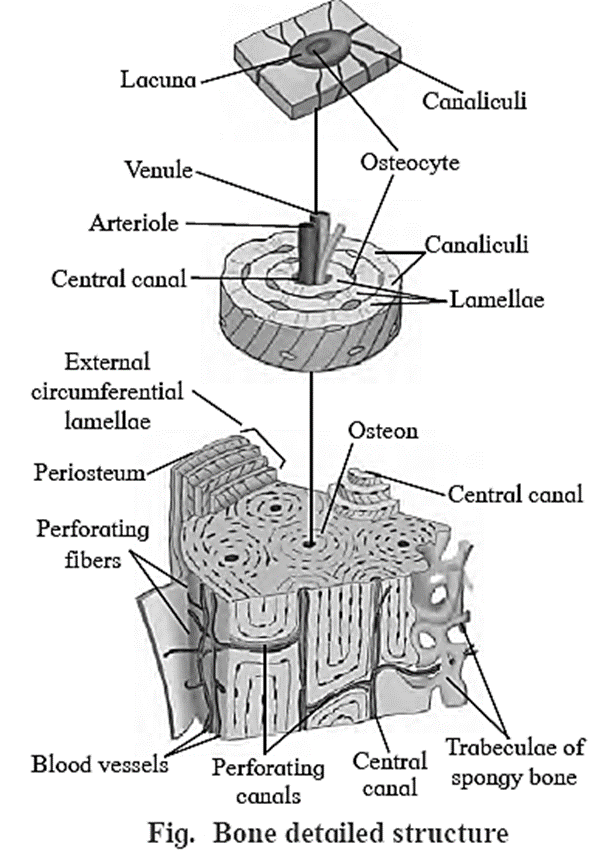
Bone :
- Protein Ossein is present in the bone matrix.
- Bone is rigid, non — pliable, dense connective tissue characterised by the hard matrix called ossein (made up of calcium salt hydroxyapatite).
- An outer tough membrane called periosteum encloses the matrix. The matrix is arranged in the form of concentric layers called lamellae.
- Bones are well vascularized and possess blood vessels and nerves that pierce through the periosteum.
- Cartilage is a supportive connective tissue. On comparison with bones, cartilage is thin, avascular and flexible. Hence, a bone is stronger than a cartilage.
Based on the presence of matrix there are two types of bones present in the human body
Spongy bones:
- Haversian system is absent in these bones.
- Rectangular matrix is arranged in the form of trabeculae.
- It contains red bone marrow.
Compact bones:
- Matrix of these bones shows haversian system without any space between the lamellae.
Functions of bone :
Bones support and protect different organs and help in movement.
Histological structure of mammalian bone :
- The bone is characterised by hard matrix called ossein which is made up of mineral salt hydroxyl apatite.
- An outer tough membrane called periosteum encloses the matrix.
- Blood vessels and nerves pierce through the periosteum.
- The matrix is arranged in the form of concentric layers called lamellae.
- Each lamella contains fluid filled cavities called lacunae from which fine canals called canaliculi radiate.
- The canaliculi of adjacent lamellae connect with each other as they traverse through the matrix.
- Active bone cells called osteoblasts and inactive bone cells called osteocytes are present in the lacunae.
- The mammalian bone shows the peculiar haversian system.
- The haversian canal encloses an artery, vein and nerves.
(D) Fluid Connective tissue (Vascular) :
Blood and lymph are fluid connective tissue present in the body of an animal
We reply to valid query.