|
Topics to be Learn :
- Introduction
- Cell cycle
- M-phase or period of division
- Types of cell division
- Amitosis
- Mitosis
- Cytokinesis
- Significance of mitosis
- Death of cell
- Meiosis
- First meiotic division or Heterotypic division (Meiosis I)
- Second meiotic division or Homotypic division (Meiosis II)
- Significance of Meiosis
|
Introduction :
Healing of wound :
- Wound is an injury to living tissue.
- Healing of wound take place by mitosis.
- Repetitive mitotic divisions near the site of injury results in healing of wound
Two types of wound-healing processes can occur, depending on the depth of the injury.
- Epidermal wound healing occurs in wounds that affect only the epidermis
- Deep wound healing occurs in wounds that penetrate the dermis.
Skin damage initiates a sequence of events that helps in repairing the skin to its normal or near-normal structure and function.
Growth : Living things show growth from within. Non-living things show growth by accumulation of materials on their surface.
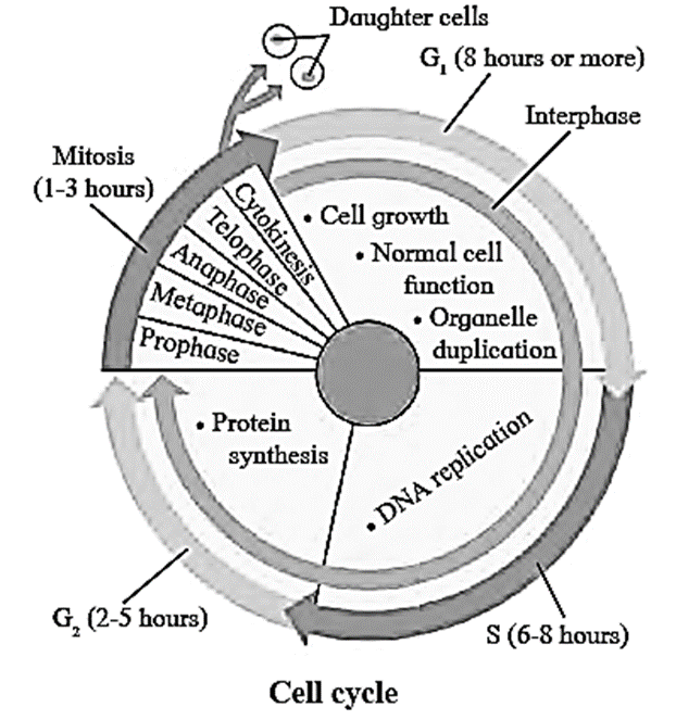
Cell cycle:
Cell cycle :
- Sequential events occurring in the life of a cell is called cell cycle.
- Interphase and M - phase are the two phases of cell cycle.
- Cell undergoes growth or rest during interphase and divides during M — phase
Click here to View Figure-1
[collapse]
Interphase: It is the stage between two successive cell divisions.
- It is the longest phase of a cell cycle.
- During interphase, the cell is metabolically very active.
- In this phase, a cell grows to its maximum size, chromosomal material (DNA and histone proteins) duplicates and the cell prepares itself for next mitotic division.
- Hence, interphase is known as preparatoiy phase.
Sub-phases of interphase:
The interphase is subdivided into three sub-phases as G1-phase, S-phase and G2-phase.
G1- phase (First gap period/First Gap Phase):
- It begins immediately after cell division.
- RNA (mRNA, rRNA and tRNA) synthesis, protein synthesis and synthesis of membranes take place during this phase.
S - phase (Synthesis phase):
- In this phase DNA is synthesized (replicated), so that amount of DNA per cell doubles.
- Synthesis of histone proteins takes place in this phase.
- If the initial amount of DNA is denoted as 2C then it increases to 4C. However, there is no increase in the chromosome number; if the cell had diploid or 2n number of chromosomes at G1, even after S phase the number of chromosomes remains the same, i.e., 2n.
- DNA replication begins in the nucleus, and the centriole duplicates in the cytoplasm,
G2- phase (Second growth phase/Second Gap Phase):
- Metabolic activities essential for cell division occur during this phase.
- Various proteins which are necessary for the cell division are also synthesized in this phase.
- Apart from this, RNA synthesis also occur during this phase.
- In animal cells, a daughter pair of centrioles appear near the pre-existing pair.
During all three phases of interphase, a cell grows by producing proteins and cytoplasmic organelles such as mitochondria and endoplasmic reticulum.
[collapse]
M-phase or period of division : 'M' stands for mitosis or meiosis.
- M-phase involves karyokinesis and cytokinesis.
- Karyokinesis is the division of nucleus into two daughter nuclei.
- Cytokinesis is division of cytoplasm resulting in two daughter cells.
Know this :
- Human cells divide once in approximately every 24 hours.
- The duration of cell cycle varies from organism to organism and from cell type to cell type.
- For e.g. yeast cell progress through cell cycle only about 90 minutes.
- In the 24 hour average duration of cell cycle of a human cell, cell division proper lasts for only about an hour. The interphase lasts more than 95% of the duration of cell cycle.
|
Go phase :
G0 phase :
- G0 is the phase of the cell cycle in eukaryotes in which many cell types stop dividing.
- If cells are deprived of appropriate growth factors, they stop at the G1 checkpoint of the cell cycle.
- Their growth and division are arrested and they remain in G0 phase.
- Mature neurons and muscle cells remain in G0 phase.
G0 phase is considered as either an extended G1 phase in which the cell is neither dividing nor preparing to divide or a distinct quiescent stage which occurs outside the cell cycle. Cells in G0 are in non-dividing phase.
[collapse]
Types of cell division :
Cell division : The division of cells into two (or more) daughter cells with same (or different) genetic material is called cell division.
Three types of cell divisions :
There are three types of cell divisions:
(i) Amitosis :
- It is the simplest form of cell division. The nucleus elongates and a constriction appears along its length
- This constriction deepens and divides nucleus into two daughter nuclei followed by division of cytoplasm resulting in formation of two daughter cells.
- This type of division is observed in unicellular organisms, abnormal cells, old cells and in foetal membrane cells.
(ii) Mitosis:
- In this type of cell division, the cell divides and form two similar daughter cells which are identical to the parent cell.
- It is completed in two steps as karyokinesis and cytokinesis.
(iii) Meiosis:
- In this type of cell division, the number of chromosomes is reduced to half. Hence, this type of cell division is also called reductional division.
- Meiosis produces four haploid daughter cells from a diploid parent cell.
[collapse]
Karyogram or Karyotype :
- A karyotype is a representation of condensed chromosomes arranged in pairs.
- Analysis o the karyotype of a particular individual indicates whether the individual has a normal set of chromosomes or whether there are abnormalities in number or appearance of individual chromosomes.
Karyokinesis :
Karyokmesis is the nuclear division which is divided into prophase, metaphase, anaphase and telophase.
Phases of Karyokinesis :
Prophase :
- In this phase, condensation of chromatin material, migration of centrosomes, appearance of mitotic apparatus and disappearance of nuclear membrane takes place.
- Due to condensation, each chromosome with its sister chromatids connected by centromere is clearly visible under light microscope.
- The nucleolus starts to disappear. ,
- Centrosome start moving towards the opposite poles of the cell.
- Mitotic apparatus is almost completely formed.
Click here to View Figure-2
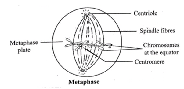
Metaphase:
- Chromosomes are completely condensed and appear short.
- Centromere and sister chromatids become very prominent.
- All the chromosomes are arranged at equatorial plane of cell. This is called metaphase plate.
- Mitotic spindle is fully formed in this phase.
- Centromere of each chromosome divides into two, each being associated with a chromatid.
[Note: The centromeres divide at the beginning of anaphase so that the two chromatids of each chromosome become separated from each other.]
Click here to View Figure-3
Anaphase:
- In this phase, chromatids of each chromosome separate and form two chromosomes called daughter chromosomes.
- The chromosomes which are formed are pulled away in opposite direction by spindle apparatus.
- Anaphase ends when each set of chromosomes reach at opposite poles of the cell.
Click here to View Figure-4
Telophase :
- This is the final stage of karyokinesis.
- The chromosomes with their centromeres begin to uncoil at the poles.
- The chromosomes lengthen and lose their individuality.
- The nucleolus reappears and the nuclear membrane appear around the chromosomes. '
- Spindle fibres breakdown and get absorbed in the cytoplasm. Thus, two daughter nuclei are formed.
Click here to View Figure-5
[collapse]
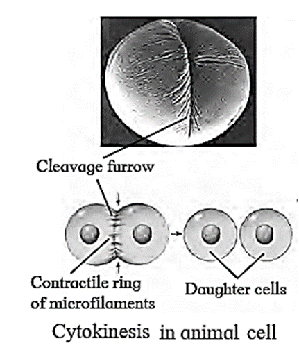
Cytokinesis :
Cytokinesis in animal cell :
Cytokinesis in animal cell :
Click here to View Figure-6
- Cytokinesis occurs by furrowing of plasma membrane that deepens and the daughter cells are formed.
- This step takes place at the end of karyokinesis (nuclear division) of mitosis.
- This process of division of cytoplasm is perpendicular to the spindle.
- It depicts the division of the cytoplasmic material in order to form two daughter cells that resemble each other.
- At the time of cytoplasmic division, organelles like mitochondria and plastids get distributed between the two daughter cells.
- Cleavage is present in this process.
[collapse]
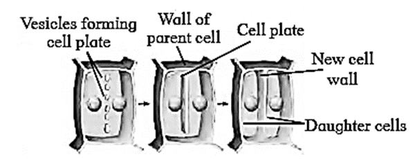
Cytokinesis in plant cell :
Cytokinesis in plant cell :
Click here to View Figure-7
- Cytokinesis occurs by the formation and extension of cell plate from center to the periphery.
- Cleavage is absent in this process.
- The formation of the new cell wall begins with the formation of a simple precursor, called the ‘cell-plate’ that represents the middle lamella between the walls of two adjacent cells.
[collapse]
Significance of mitosis :
Significance of mitosis :
- As mitosis is equational division, the chromosome number is maintained constant.
- It ensures equal distribution of the nuclear and the cytoplasmic content between the daughter cells, both quantitatively and qualitatively. Therefore, the process of mitosis also maintains the nucleo-cytoplasmic ratio.
- The DNA is also equally distributed.
- It helps in growth and development of organisms.
- Old and worn-out cells are replaced through mitosis.
- It helps in the asexual reproduction of organisms and vegetative propagation in plants.
- Through mitotic divisions, meristematic tissues such as the apical and lateral cambium show continuous growth of plants throughout their life.
[collapse]
Life span of cell :
Life span of cell :
- Life span of different cells vary greatly.
- Life span of a cell is decided by its growth rate, metabolic activities and cell size.
- The life span of a cell can be analysed in laboratory by applying carbon-14 technique to DNA. This method is commonly used in archaeology and paleontology to find the age of fossils. Same can be applied to determine the life span of a cell.
[collapse]
Death of cell :
Necrosis : Necrosis is a form of cell injury which leads to the premature death of cells. For example: due to scrape or a harmful chemical.
Apoptosis : Apoptosis also known as programmed cell death or cellular suicide. In apoptosis cells die in controlled way.
- Example: during embryonic development the cells between the embryonic fingers die in a process called apoptosis to give a definite shape to the fingers.
- Apoptosis also helps in eliminating potential cancer cells.
Meiosis :
- It takes place only in reproductive cells during the formation of gametes.
- By this division, the number of chromosomes is reduced to half, hence it is also called reductional division.
- The cells in which meiosis take place are termed as meiocytes.
- Meiosis produces four haploid daughter cells from a diploid parent cell.
Meiosis is of two subtypes :
(i) First meiotic division or Heterotypic division – (Meiosis I) :
- Heterotypic division is first meiotic division, during which a diploid cell is divided into two haploid cells
- The daughter cells resulting from this division are different from the parent cell in chromosome number.
- Hence the division is called heterotypic division. .
It consists of following phases:
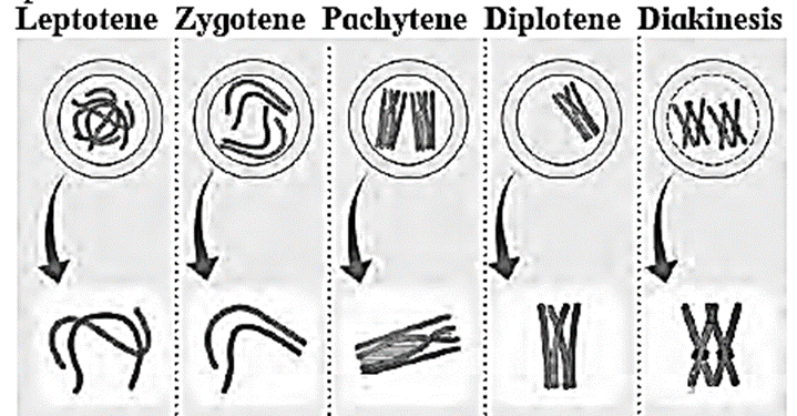
Prophase -I:
- It is the most complicated and longest phase of meiotic division
- It is further divided into five sub-phases viz. leptotene, Zygotene, pachytene, diplotene and diakinesis.
Sub-phases of Prophase-I :
Click here to View Figure-8
Leptotene :
- The volume of the nucleus increases.
- The chromosomes become long distinct and coiled.
- They orient themselves in a specific form known as bouquet stage. This is characterized with the ends of chromosomes converged towards the side of nucleus where the centrosome lies.
- The centriole divides into two and migrate to opposite poles,
Zygotene :
- Pairing of non-sister chromatids of homologous chromosomes takes place by formation of synaptonemal complex. This pairing is called synapsis.
- Each pair consist of a maternal chromosome and a paternal chromosome.
- Chromosomal pairs are called bivalents or tetrads.
Pachytene:
- Each individual chromosome begins to split longitudinally into two similar chromatids.
- Therefore, each bivalent now appears as a tetrad consisting of four chromatids.
- The homologous chromosomes begin to separate but they do not separate completely and remain attached to one or more points.
- These points are called chiasmata (Appear like a cross-X).
- Chromatids break at these points and broken segments are exchanged between non-sister chromatids of homologous chromosomes resulting in recombination.
Diplotene :
- The chiasmata become clearly visible in diplotene due to beginning of repulsion between synapsed homologous chromosomes. This is known as desynapsis.
- Synaptonemal complex also starts to disappear.
Diakinesis :
- The chiasmata beings to move along the length of chromosomes from the centromere towards the ends of chromosomes. The displacement of‘ chiasmata is termed as terminalization.
- The terminal chiasmata exist till the metaphase.
- The nucleolus and nuclear membrane completely disappear and spindle fibres begin to appear. .
[collapse]
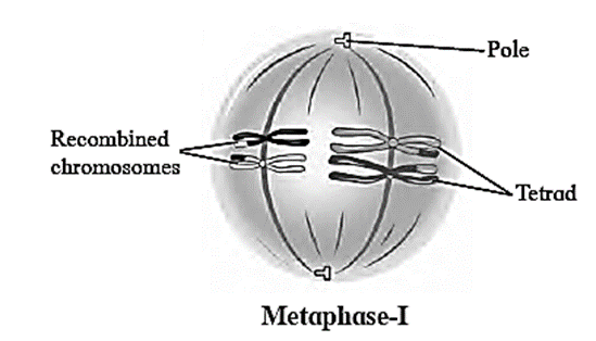
Metaphase-I :
- The spindle fibres are well developed.
- The tetrads orient themselves on equator in such a way that centromeres of homologous tetrads lie towards the poles and arms towards the equator.
- They are ready to separate as repulsive force increases.
Click here to View Figure-9
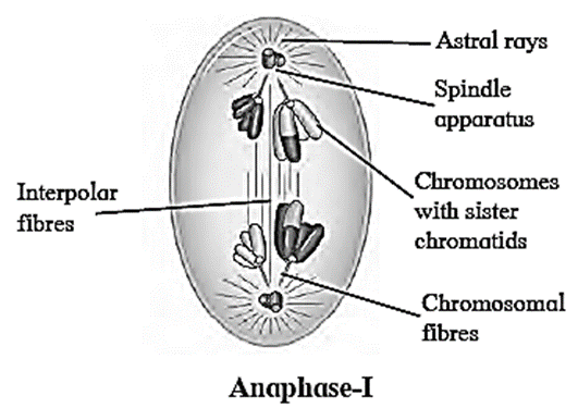
Anaphase-I:
- Homologous chromosomes are carried towards the opposite poles by spindle apparatus. This is known as disjunction.
- The two sister chromatids of each chromosome do not separate in meiosis -I. This is reductional division.
- The sister chromatids of each chromosome are connected by a common centromere.
- Both sister chromatids of each chromosome are now different in genetic content as one of them has undergone recombination.
Click here to View Figure-10
Telophase-I: 4
- The haploid number of chromosomes becomes uncoiled and elongated after reaching their respective poles.
- The nuclear membrane and nucleolus reappear and thus two daughter nuclei are formed.
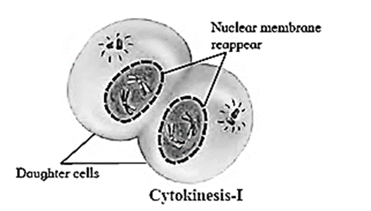
Cytokinesis-I :
- After the karyokinesis, cytokinesis occurs and two haploid cells are formed.
- In many cases, these daughter cells pass through a short resting phase or interphase / interkinesis.
Click here to View Figure-11
The association between the homologous chromosomes i.e. chiasmata remain till metaphase I. During metaphase I, the paired homologous chromosomes move to the metaphase plate. In anaphase I, the spindle fibers begin to shorten. As these spindle fibres shorten, the association between homologous chromosomes (chiasmata) are broken, allowing homologous chromosomes to be pulled to opposite poles.
Q. What is exact structure of synaptonemal complex.
Answer :
Synaptonemal complexes are zipper like structures assembled between homologous chromosomes during the prophase of first meiotic division.
[collapse]
Q. What is structure of chiasmata?
Answer :
Chiasmata are X-shaped points of attachment between two non-sister chromatids of a homologous chromosomes.
[collapse]
Spindle fibres :
Spindle fibres :
- Spindle fibres are formed from microtubules with many accessory proteins.
- Spindle fibres elongate for assembly of chromosomes at equatorial plane of the cell during metaphase and spindle fibres contract for pulling chromosomes towards opposite poles during anaphase.
- The spindle fibres elongate (polymerize) by incorporating subunits of the protein tubulin and contract (depolymerize) by losing subunits.
[collapse]
Role of Centriole : Centriole plays an important role in cell division. Centrioles help organize icrotubule assembly and forms spindle apparatus that separate the chromosomes during cell division.
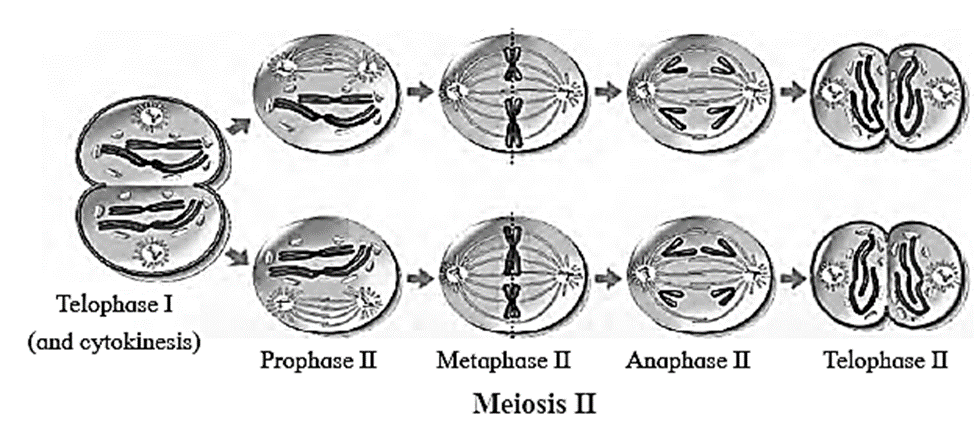
(ii) Second meiotic division or Homotypic division (Meiosis II) :
Two haploid cells formed during first meiotic division divide further into four haploid cells this division is called homotypic division.
It consists of five phases: prophase - II, metaphase — II, anaphase — II, telophase - II, and Cytokinesis — II. .
Five phases of Meiosis-II :
Click here to View Figure-12
Prophase — II:
- The chromosomes are ‘distinct with two chromatids.
- Each centriole divides into two resulting in formation of two centrioles which migrate to opposite poles and form asters.
- Spindle fibres are formed between the centrioles.
- The nuclear membrane and nucleolus disappears in this phase.
Metaphase — II:
- Chromosomes are arranged at the equator.
- The two chromatids of each chromosome are separated by division of the centromere.
- Some of the spindle fibres are attached to the centromeres and some are arranged end to end between two opposite centrioles.
Anaphase — II:
In this phase, the separated chromatids become daughter chromosomes and move to opposite poles due to the contraction of the spindle fibres attached to centromeres.
Telophase — II:
- In this stage daughter chromosomes starts to uncoil.
- The nuclear membrane surrounds each group of chromosomes.
- Nucleolus reappears in this phase.
Cytokinesis — II
- Cytokinesis takes place after the nuclear division.
- Two haploid cells are fonned from each haploid cell.
- Thus, four haploid daughter cells are formed.
- These cells then undergo changes to form gametes.
[collapse]
Significance of Meiosis :
- Meiotic division produces gametes.
- If it is absent, the number of chromosomes would double or quadruple resulting in the formation of monstrosities (abnormal gametes).
- Meiosis ensures that organisms produced by sexual reproduction contain correct number of chromosomes.
- Meiosis exhibits genetic variation by the process of recombination.
- Variations increase further after union of gametes during fertilization creating offspring with unique characteristics.
- Thus, it creates diversity of life and is responsible for evolution.
In absence of meiosis :
- Production of gametes will not take place. Gametes are essential for sexual reproduction, which are produced by the process of meiosis
- Diploid organisms have two set of chromosomes (one paternal and one maternal).
- For a diploid organism to undergo sexual reproduction it needs to create gametes that contain only one set of chromosomes so the number of chromosomes remains same in the next generation.
- In absence of meiosis, the chromosome number of parents and their offspring will differ in every generation, hence no species will hold its characters.
- Also, there will be no crossing over of homologous chromosomes. Thus, there will be no variations with respect to the changing environment in progeny to maintain their existence, which may lead to extinction of species.
Difference between meiosis — I and meiosis — II :
Difference between meiosis — I and meiosis — II :
| Meiosis — I |
Meiosis — II |
| 1-Diploid cell is divided into two haploid cells.
2-This division is called heterotypic division.
3-It consists of prophase-I, metaphase-I, anaphase-I, telophase-I and cytokinesis.
4-Number of chromosomes is reduced to half, i.e. from diploid to haploid state.
5-It is complicated and long duration division.
6-Telophase I results into 2 daughter cells.
|
1-Two haploid cells formed in meiosis I divides further into four haploid cells
2-This division is called homotypic (equational) division.
3-It consists of prophase — II, metaphase —II, anaphase — II, telophase — II and cytokinesis.
4-In meiosis II number of chromosomes remain the same.
5-It is simple and short duration division, Telophase II results in 4 daughter cells.
|
[collapse]
Process of recombination :
Process of recombination :
- Recombination is exchange of genetic material between paternal and maternal chromosomes during gamete formation.
- The points where crossing over takes place is known as chiasmata.
- Chromatids acquire new combinations of alleles by physically exchanging segments in crossing-over.
- The exchange of genetic material between homologous chromosomes involves accurate breakage an joining of DNA molecules through a complex mechanism.
- It is catalyzed by enzymes.
[collapse]


We reply to valid query.