Notes
|
Topics to be Learn :
|
Introduction :
Types of animals :
Animals on earth show great diversity. The different types of animals present around us are
- Unicellular and multicellular
- Prokaryotic and eukaryotic
- Veitebrates and invertebrates
- Unisexual and hennaphrodite
- Aquatic, terrestrial, amphibian, reptilian, aerial, etc.
Habit and Habitat :
- Cockroaches are said to be omnipresent as they are present everywhere, all over the world.
- They are usually seen in damp and moist places, crevices.
- Cockroaches are active at night hence, they are termed as nocturnal.
- They are cursorial insects as they show terrestrial adaptations for running
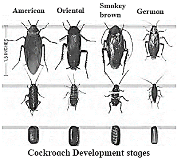
- Common species of cockroach found in India are Periplanera americana, Blarta orientalis and Blarra germanica
- Cockroaches exhibit cannibalism. It is the tendency of an organism to eat its young one as food.
Systematic Position :
| Classification | Reason |
| Kingdom : Animalia | Cell wall absent, heterotrophic nutrition.) |
| Phylym : Arthropoda | Jointed appendages are present, segmented body, chitinous exoskeleton. |
| Class : Insecta | Two pairs of wings and three pairs of walking legs are present. |
| Genus : Periplaneta | Nocturnal, straight wings. |
| Species : americana | Origin is in Continent of America |
External Morphology :
External morphology of cockroach:
Shape and size: Cockroach has elongated, bilaterally symmetrical, dorso-ventrally flattened and truly segmented body. They are tiiploblastic and eucoelomate. The body cavity called haemocoel is filled with the fluid haemolymph.

Coloration: Their colour is glistening brown or reddish ‘brown.
Exoskeleton: Tough, waxy, non-living chitinous exoskeleton protects the body of the cockroach. It is made up of nitrogenous polysaccharide - chitin that provides strength, elasticity and surface area for attachment of muscles. Each body segment of cockroach is covered by four chitinous plates called sclerites namely, dorsal tergum, ventral stemum and two lateral pleurons.
Body division: The body is divided into three regions viz. head, thorax and abdomen.
Head: It is fonned by the fusion of six segments. It is triangular or ovate in shape. The head is highly mobile due to flexible neck. It bears a pair of long antennae, a pair of compound eyes and mouthparts adapted for biting and chewing of food.
Head of cockroach bears following ‘structures:
(i) Antennae:
- They are also called as feelers.
- These are long, filamentous, segmented structures that can move in all directions.
- They are lodged in membranous pits known as antenna] sockets.
- They are tactile (touch) as well as olfactory (smell) organs.

- Function: They are useful in ocating the food material in the vicinity.
(ii) Fenestrae: Fenestrae also called as ocellar spots. They are situated at the base of each antenna and appears as white spots.
(iii) Compound eyes:
- Compound eyes are paired, dark, kidney-shaped structures located on the head's dorsolateral sides.
- They are composed of a large number of hexagonal ommatidia, approximately 2000 ommatidia (sing. ommatidium). These ommatidia are the structural and functional units of the compound eye, each of which forms an image of a very small portion of the visual field.
- Collectively, the compound eye produces a mosaic image. Even though the compound eye gives a mosaic or hazy vision yet the animal can detect the slightest movement of the object.
- Compound eyes provide low resolution and more sensitive vision.
(iv) Mouthparts:
- Cockroach has a pre-oral cavity in front of the mouth in which food is received. It is bounded by mouthparts which are of chewing and biting type.
- This includes: labrum, labium, a pair of mandibles, a pair of maxillae, tongue like hypopharynx (present at the centre of mouth).
- Salivary duct opens at the base of hypopharynx and the mouth opens into foregut.

Thorax: Thorax is made up of three distinct segments - prothorax (anterior segment), mesothorax and metathorax (posterior segment). Ventrally, the thorax bears three pairs of walking legs, one at each segment. Dorsally, the thorax bears two pairs of wings attached to mesothoracic and metathoracic segment of the body.
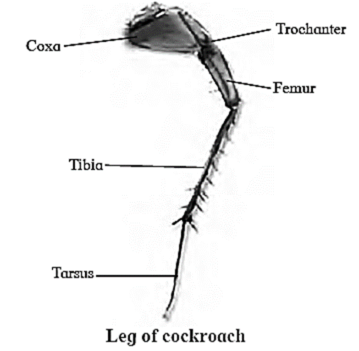
- Legs: On the ventral side, there are three pairs of walking legs. Each leg has five podomeres: coxa, trochanter, femur, tibia, and tarsus. Tarsus is the final segment, which is made up of five movable segments called tarsomeres. The final tarsomere has a pair of claws and a cushion—like arolium—that aid in clinging.
- Wings: Forewings and hindwings form the two pair of wings present on the dorsal side. Forewings are the first pair of dark, opaque, thick, leathery wings. Hindwings are thin, broad, membranous delicate and transparent.They are attached to tergum of metathorax. Functions: Forewings are protective in function and hindwings are helpful in flight, thus are called true wings. ,
Abdomen:
- The abdomen is elongated and made up of ten segments. Each segment has a dorsal tergum and ventral stemum.
- Tergum is jointed to the stemum laterally by a soft cuticle called pleura
- The posterior segments are telescoped in. Due to this, the eighth and ninth terga get overlapped by the seventh. The tenth tergum projects backward and is deeply notched. It bears a pair of small, many jointed anal cerci.
- Anal cerci are a pair of appendages at the end of the abdomen, that arise from the 10th segment of the body of cockroach.
- The abdomen is narrow and tapering in. males as compared to females. The ninth stemum of males also bears a pair of short, unjointed anal style.
Spiracles: These are series of slit-like openings on either side of the body. There are ten pairs in all, two on thorax and eight on abdomen. The spiracles let the air into and out of the tubes called trachea.
Know This :
|
Body cavity :
- Around the viscera is a body cavity or true coelom.
- The body cavity is filled with blood due to the open type of circulation. As a result, it is known as haemocoel.
- Fat bodies can be seen in the haemocoel. It appears as a loose, whitish mass of tissue.
- The fat body is composed of large, polygonal cells that contain fat globules, proteins, and occasionally glycogen.
Digestive System of Cockroach :
Digestive system of cockroach consists of mouthparts, alimentary canal and salivary glands.

Mouth parts : Pre-oral cavity present in front of the mouth receives food. It is bounded by chewing and biting type of mouth parts. These are movable, segmented appendages that help in ingestion of food.
The mouthparts of cockroach comprises of:
Labrum: It forms the upper lip. It is a single flap-like movable part which covers the mouth from upper side. It forms an anterior wall of pre-oral cavity.
- Function : It is useful in holding the food during feeding.
Mandibles: These are two dark, hard, chitinous structures with serrated median margins. They are true jaws present on either side, behind the labrum.
- Function: They perform co-ordinated side-wise movements with the help of adductor and abductor muscles to cut and crush the food.
Maxillae: These are the accesssory jaws. They are also called as first pair of maxillae. These are situated on the either side of mouth behind the mandibles. Each maxilla consists of sclerites like cardo, stipes, galea, lacinia and maxillary palps.
- Functions: Maxillae hold food, help mandibles for mastication. They are also used for cleaning the antennae and front legs. Maxillary palps act as tactile organs.
Labium: It forms the lower lip. Labium is also known as second maxilla which covers the pre-oral cavity from the ventral side. It is firmly attached to the posterior part of head. It has three jointed labial palps which are sensory in function.
- Function: It is useful in pushing the chewed food in the pre—oral cavity. It prevents the loss of food falling from the mandibles, while chewing.
Hypopharynx : Hypopharynx / tongue / lingua is a somewhat cylindrical single structure, located in front of the labium and between first maxillae. A salivary duct opens at the base of Hypopharynx. Hypopharynx bears comb-like plates called super-lingua on either side.
- Function: It is useful in the process of feeding and mixing saliva with food.
Alimentary canal: It is long (6 - 7cm) tube of different diameters and two openings
Cockroach does not have the buccal cavity thus, the alimentary canal begins from pre-oral cavity
The alimentary canal is divisible into three parts: foregut, midgut and hindgut.
(i) Foregut or stomodaeum : It consists of pharynx, oesophagus, crop and gizzard.
- Pharynx: It is very short, narrow but muscular tube that opens into oesophagus. It containstaste sensillae. Function: Conduction of food into the oesophagus
- Oesophagus: It is slightly long and narrow tube which opens into crop.
- Crop: Crop is a large, pear shaped and sac-like organ. Function: It temporarily stores the food and then sends it to gizzard.
- Gizzard: Gizzard or proventriculus is a small spherical organ. It is provided internally with a circlet of six chitinous teeth and backwardly directed bristles. The foregut ends with gizzard. Function: The chitinous teeth present in gizzard are responsible for crushing the food and the bristles help to filter the food.

(ii) Midgut or mesenteron: It consists of stomach and hepatic caeca.
- Ventriculus or stomach: It is straight, short and narrow. Stomach is lined by glandular epithelium which secretes digestive enzymes. It is mainly responsible for digestion and absorption.
- Hepatic caeca: These are thin, transparent, short, blind (closed) and hollow tubules. Function: They secrete digestive enzymes.
(iii) Hindgut or proctodaeum : It consists of ileum, colon and rectum.
- Ileum: It is short and narrow part of hindgut. Malpighian tubules open in the anterior lumen of ileum, near the junction of midgut and hindgut. Posterior region of ileum contains sphincter. Ileum directs the nitrogenous wastes and undigested food towards colon.
- Colon: It is a longer and wider part of the hindgut. It directs waste material towards the rectum. It reabsorbs water from wastes as per the need.
- Rectum: It is oval or spindle-shaped, terminal part of the hindgut. It contains six rectal pads along the internal surface for absorption of water. Rectum open into anus. Anus is present onthe ventral side of the 10th segment. It is the last or posterior opening of the digestive system. The undigested food is released out of the body through anus.
Salivary glands:
- Cockroach has a pair of salivary glands which secrete saliva.
- Each salivary gland has two glandular lobes and a receptacle or reservoir.
- The glandular lobes consists of several irregular-shaped white coloured lobules which secrete saliva.
- Each gland has a salivary duct. Both the ducts unite to form a common salivary duct.

- Receptacle of each salivary gland is thin-walled, elongated, sac-like structure. Each receptacle has a duct. These ducts unite to form common reservoir duct.
- Common salivary duct and common reservoir duct unite together to form a common efferent salivary duct. The efferent salivary duct opens at the base of tongue or hypopharynx.
Circulatory System or Blood Vascular System :
Cockroach has open circulatory system. It consists of colourless blood (haemolymph), a dorsal blood vessel (heart and dorsal aorta) and haemocoel.
Haemolymph: Haemolymph is colourless as it is without any pigment. It consists of plasma and seven types of blood cells/haemocytes. Plasma consists of water with some dissolved organic and inorganic solutes. It is rich in nutrients and nitrogenous wastes like uric acid.
Haemocoel: The body cavity of cockroach (haemocoel) can be divided into three sinuses due to two diaphragms i.e. dorsal and ventral diaphragm. These diaphragms are thin, fibromuscular septa (sing.septum). It remains attached to terga along lateral sides at intermittent points.
(i) Dorsal diaphragm : It has 12 pairs (10 abdominal and 2 thoracic) of fan-like alary muscles. Alary muscles are triangular with pointed end attached to terga at lateral side and broad end lies between the heart and dorsal diaphragm.
(ii) Ventral diaphragm: It is flat and present just above the ventral nerve cord. Laterally, it is attached to stema at intermittent points.
(iii) Sinuses: The coelom of cockroach is divided into three sinuses — pericardial sinus, perivisceral sinus and perineural sinus.
- Pericardial sinus : It is dorsal, very small and contains dorsal vessel. _
- Perivisceral sinus : It is middle and largest sinus. It contains fat bodies and almost all major visceral organs of alimentary canal and reproductive system.
- Perineural sinus : It is ventral, small and contains ventral nerve cord. It is continuous into legs. All the three sinuses communicate with each other through the pores present between two successive points of attachments of diaphragms.
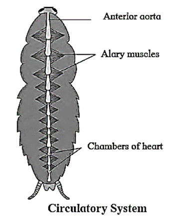
Dorsal blood vessel : This is present in pericardial sinus, just below the tergum. It is divisible into posterior heart and anterior aorta (dorsal aorta/cephalic vessel). _ .
- Heart : It is about 2.5 cm long, narrow, muscular tube that is open anteriorly and closed posteriorly. It starts from 9th abdominal segment and extends anteriorly upto 1st thoracic segment. Heart of cockroach is 13 chambered, out of which 10 chambers are in abdominal region and three are in thoracic region. Each chamber has a pair of vertical slit-like incurrent aperture or opening called ostium (plural: ostia). Ostia are present along lateral side in the posterior region of first 12 chambers;Each ostium has lip-like valves that allow the flow of blood from sinus to heart only.
- Anterior aorta : Heart is continued by a short, thin-walled vessel called dorsal aorta. It lies in head region and opens in haemocoel.
.
Mechanism of blood circulation:
- Blood (haemolymph) circulates between sinuses and heart due to contraction and relaxation of heart and alary muscles.
- The heart contracts (systole) and relaxes (diastole) alternatively. After diastole, there is a third phase in the heart cycle known as diastasis. During diastasis, heart remains in expanded state.
- During diastole, alary muscles contract, making the dorsal diaphragm flat. As a result, blood passes from perivisceral sinus to pericardial sinus through fenestrae and finally to the heart through ostia. i
- During systole, contraction starts at the posterior end and the wave of contraction passes anteriorly. Due to this, blood is pushed towards the dorsal aorta.
- Ostia remain closed with the help of valves, during systole. As a result, blood flushes into head region from where it goes to perineural sinus and then to perivisceral sinus.
- During systole, alary muscles are relaxed and due to this, the dorsal diaphragm becomes convex.
- The volume of pericardial sinus is now reduced. This makes the blood to move from pericardial sinus to perivisceral sinus through fenestrae.
Know This :
|
Respiratory System or Tracheal System :
Cockroaches have an internal respiratory system of air tubes called the tracheal system, which brings air into the body and makes contact with every pan. It enables direct gas exchange between air and tissues without the use of blood.
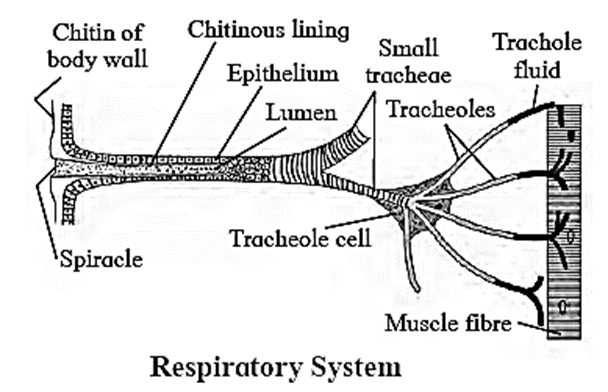
These air tubes of internal respiratory system begin at the opening on body surface called spiracles.
- Spiracles: They are paired respiratory openings. Spiracles are present on the ventro-lateral side of the body, of thoracic and eight pairs of abdominal spiracles. The in pleural membrane. Cockroaches have two pairs spiracles open into a series of air sacs from which arise the tubes called trachea.
- Trachea: The trachea form a definite pattern of branching tubes arranged transversely as well as longitudinally. They are about 1 mm thick and have spiral or annular thickening of chitin. The inner lining chitin prevents the trachea from collapsing. Each trachea further branches into smaller tubes called tracheoles.
- Tracheoles : These are fine intracellular tubes that penetrate deep into tissues. They are thin and not lined by chitin. They end blindly in the cells. Each tracheole at the blind end is filled with a watery fluid through which exchange of gases takes place. The content of the fluid keeps changing. At high muscular activity, part of fluid part is drawn into the tissues to enable more and rapid oxygen intake.
- The rhythmic movements of thoracic and abdominal muscles are involved in the renewal of air in tracheal system.
- The thin branching tubes (tracheal tubes subdivided into tracheoles) carry oxygen from the air to all the parts.
- The opening of spiracles is regulated by sphincters.
Excretory System :
- Malpighian tubules are the main excretory organs of cockroach.
- They are thin, yellow coloured, ectodermal thread-like structures that lie in the haemocoel.
- These tubules are 150 in number. Malpighian tubules are attached to the alimentary canal between the midgut and hindgut.
- Each Malpighian tubule is lined with a single layer of glandular epithelial cells having microvilli. The distal portion of Malpighian tubule is secretory and the proximal part is absorptive in function.
- They extract water and nitrogenous wastes from the haemocoel and convert them into uric acid and pass them into ileum. As the cockroach excretes uric acid, it is said to be uricotelic.
- Also, fat bodies, nephrocytes and uricose glands (only in males) help in excretion.
- In cockroach, nephrocytes (urate cells) associated with fat bodies and cuticle are also believed to be excretory in function. The nephrocytes are cells present along with the fat bodies or present along the heart and store nitrogenous wastes.
- The excretory products later are removed in the haemocoel. Some nitrogenous wastes are deposited on the cuticle and eliminated during moulting.
Nervous System :
Nervous system of cockroach is ventral, solid and ganglionated. It consists of central nervous system (CNS), peripheral nervous system (PNS) and autonomous nervous system (ANS).
- The nervous system of cockroach is spread throughout the body.
- The head of cockroach holds a bit of nervous system while the rest is situated along the ventral part of its body.
Central nervous system (CNS): Central nervous system consists of (1) nerve ring and (2) ventral nerve cord.

(1) Nerve ring consists of:
- a pair of supra-oesophageal ganglia
- a pair of circum-oesophageal connectives
- a pair of sub-oesophageal ganglia
(i) Supra-oesophageal ganglia or cerebral ganglia : A pair of supra-oesophageal ganglia is collectively known as the brain. Brain is present in head, above the oesophagus and between antennal bases. Each supra-oesophageal ganglion is formed by the fusion of three small ganglia - protocerebrum, deutocerebmm and tritocerebrum.
(ii) Circum-oesophageal connectives: Supra-oesophageal ganglia are connected to sub-oesophageal ganglion by a pair of lateral nerves called as circum-oesophageal connectives. Connectives arise from supra-oesophagial ganglia.
(iii) Sub-oesophageal ganglia: It is a bilobed and present below the 0esophagus, in head. It is also formed by the fusion of three pairs of ganglia.
(2) Ventral nerve cord :
- It arises from the sub-oesophageal ganglion. It is present along mid-ventral position, in perineural sinus.
- It is double ventral nerve cord and consists of nine segmental, paired ganglia. .
- First three pairs of segmental ganglia are large and known as thoracic ganglia. The other six pairs of segmental ganglia are in abdomen (abdominal ganglia).
- 6th abdominal ganglion is the largest and it is present in 7th abdominal segment.
- There is no ganglion in 6th segment.
Peripheral nervous system (PNS):
- The peripheral nervous system comprises of nerves that arise from various ganglia of CNS.
- Six pairs of nerves arise from the supra-oesophageal ganglia.They supply to the eyes, antenna and labrum.
- Nerves arising from the sub-oesophageal ganglion supply to the mandibles, maxillae and labium
- Nerves arising from the thoracic ganglia supply to the wings, legs and intemal thoracic organs.
- Nerves from abdominal ganglia go to the abdominal organs of respective abdominal segments.
Autonomic nervous system (ANS): It consists of four ganglia and a retro cerebral complex.
The ganglia are as follows:
- Frontal ganglion: It is present above the pharynx and in front of brain.
- Hypocerebral ganglion: It is present on the anterior region of oesophagus.
- Ingluvial ganglion: It is present on crop. It is also called as visceral ganglion.
- Ventricular ganglion: It is present on gizzard.
Ganglion is a group of nerve cell bodies.
Reproduction System :
Male reproductive system :

- Male reproductive system includes primary and secondary reproductive organs.
- Primary sex organs (male gonads) are called testes. They are paired and located in the 4th and 6th abdominal segments. Sperms produced in testis are carried by vasa deferentia.
- Vasa deferentia is a pair of thin tubular structure that arise from the testes and open into ejaculatory duct through seminal vesicle.
- They carry sperms to ejaculatory duct.
- Ejaculatory duct opens into male gonopore situated ventral to anus.
- Sperms produced by testis are pstored in seminal vesicles in the fonn of bundles called spermatophores. These spennatophores are deposited in female reproductive tract during copulation.
- Mushroom shaped gland or utricular gland is an accessory reproductive gland. It is present in the 6th – 7th abdominal segments.
- Male gonapophyses or phallomere form the external genitalia of male. These are three asymmetrical chitinous structures surrounding the male gonophore.
Female reproductive system :

- Female reproductive system consists of primary and secondary reproductive organs.
- Ovaries are primary reproductive organs. They are paired and lie lateral in position in 2nd - 6th abdominal segments. Each ovary is formed of a group of eight ovarian tubules or ovarioles, containing a chain of developing ova.
- All ovarioles of an ovary open in lateral oviduct of respective side.
- Both the lateral oviducts unite to form a common oviduct or vagina.
- Common oviduct or vagina opens into the Bursa copulatrix (genital chamber), the female organ of copulation.
Know This :
| In males, genital pouch or genital chamber lies at the hind end of abdomen which is bounded dorsally by 9th and 10th terga and ventrally by the 9th sternum. The genital pouch contains dorsal anus, ventral male genital pore and gonapophysis. | In females, the 7th sternum is boat shaped and together with 8th and 9th sternum forms a brood or genital pouch whose anterior pan contains gonopore, spennathecal pores and collateral glands. |
Fertilization and formation of ootheca :
Process of fertilization in cockroach :
- The process of fertilization in cockroach is internal.
- Male and female cockroaches come together by their posterior phallomeres.
- The spermatophores are transferred to the genital chamber of female cockroach.
- Sperms released from the spermatophore reach the spermatheca.
- The eggs are discharged from both the ovaries alternately into the common oviduct and pass into the genital chamber.
- Sperms coming from the spermatheca fertilize the eggs in the genital chamber.
Ootheca :
- The secretion of collaterial glands forms a capsule around them is called as ootheca or egg case.
- It is about 8 mm long and ranges from dark reddish to blackish brown.
- Ootheca contains 14 to 16 fertilized eggs in two rows.
- They are dropped or glued to a suitable surface, like a crack or crevice of good humidity near the food source.
- A female cockroach on an average, produces 9 to 10 oothecae during its lifespan.
Stages of development in cockroach :
- The development in cockroach (Periplaneta americana) is paurametabolous as the development occurs through nymphal stage.
Fertilized egg ——> Nymph ——> Adult
- The nymph looks like adult but it is smaller and sexually immature.
- After sufficient growth, nymph undergoes moulting and enters into a stage between two successive moults known as instar.
- Cockroaches may undergo moulting for around 13 times before reaching the adult stage.
- The nymphal stages have wing pads but only adult cockroaches have wings.
- The embryonic period in cockroach varies as per temperature and humidity. At 24°C, the duration is about 58 days and at 30° C, the duration is about 32 days.
Interactions with mankind :
Cockroaches are considered pests due to following reasons:
- Cockroach causes damage to the household materials like clothes, shoes, paper etc. They also eat and destroy the food stuff.
- They contaminate food which gives typical smell and make it unpalatable.
- As they live in sewage pipes and gutter holes, they carry with them harmful pathogens causing diseases like cholera, diarrhoea, tuberculosis, typhoid, etc.
- Cockroaches are considered as bio-indicators as their presence indicates unhygienic conditions
Cockroach serves as a part of food chain :
- Many amphibians, birds, lizards and rodents prey upon them making them a part of food chain.
- They are eaten by certain groups of people in South America, China and Myanmar.
Cockroach is used as experimental animal in laboratories and for biological research, as they can be obtained easily without causing damage to ecological balance.
Control Measures : As cockroach is economically harmful organism it must be controlled in an efficient way. Some of the measures are as follows :
- Good Sanitation : Dark and humid places of kitchen, cupboards, trolleys must be cleaned regularly. Cracks and crevices and such areas must be filled. There should not be any place in a home, where accumulation of garbage may take place.
- Keeping the drain trap filled with water : Cockroaches frequently enter home by migrating up from sewer connections if the drain trap is dry. So always keep the drain trap filled with water.
- Chemical control : Organophosphates, carbamates, pyrethroids and boric acid are efficient poisons of cockroaches, various types of their formulations are available in market, under various brand names.
We reply to valid query.