Notes-Part-2-Class-11-Science-Biology-Chapter-8-Plant Tissues and Anatomy-Maharashtra Board

Plant Tissues and Anatomy

Maharashtra Board-Class-11-Science-Biology-Chapter-8

Notes

Topics to be Learn : Part-1

  • Tissue
  • Meristematic Tissue
  • Permanent Tissue
  • Tissue Systems

Topics to be Learn : Part-2

  • Secondary Growth in Plants
  • Wood
  • Cork Cambium and Secondary Growth
  • Anatomy of Root, Stem and Leaf

Secondary growth in plants :

  • Dicotyledonous plants and gymnosperms exhibit increase in girth of root and stem.
  • In dicot stem, secondary growth begins with the formation of a continuous cambium ring.
  • The cambium present between the primary xylem and primary phloem of a vascular bundle is called intrafascicular cambium.
  • The cells of medullary rays adjoining these intrafascieular cambium strips become meristematic (regain the capacity to divide) and form the interfascicular cambium.
  • Thus, a complete and continuous ring of vascular cambium is formed.
  • The cambium ring cuts off new cells, towards both inner and outer sides.
  • The cells that are cut-off towards pith (inner side) mature into secondary xylem and cells that are cut-off towards periphery mature into secondary phloem.
  • Generally, amount of secondary xylem is more than the secondary phloem.

Formation of cambial ring :

Formation of cambial ring :

  • The cambium present between the primary xylem and primary phloem of a vascular bundle is called intrafascicular cambium.
  • With the onset of favourable season, meristematic cells of intrafascicular cambium become active.
  • Simultaneously, the ray parenchyma cells, both fusiform initials and ray initials become meristematic. This is known as dedifferentiation.
  • These form patch of cambial cells (meristematic cells) in between the adjacent bundles and produce interfascicular cambium.
  • Both intrafascicular and interfascicular cambium join and form a complete ring known as the cambial ring. This is possible because they lie in one plane.

[collapse]

Secondary growth in roots :

Secondary growth in roots :

  • Secondary growth is observed in most of the dicot and gymnospermic root by producing secondary tissue and periderm.
  • Secondary growth is produced by vascular cambium and cork cambium respectively.
  • Conjunctive parenchyma cells present on the inner edges of primary phloem bundles become meristematic.
  • These cells add secondary xylem and secondary phloem on the inner and outer side respectively which results in secondary growth.

[collapse]

Wood :

  • Growth rings are formed due to cambial activity during favourable and non-favourable climatic conditions.
  • During favourable conditions, spring wood (early wood) is formed which has broader xylem bands, lighter colour, tracheids with thin wall and wide lumen, fibres are less in number, low density. Whereas, during unfavourable conditions, autumn wood (late wood) is formed which has narrow xylem band, darker in colour, lumen is narrow and walls are thick with abundant fibres, high density.
  • Spring wood and autumn wood that appear as alternate light and dark concentric rings, constitute an annual ring or growth ring.
  • These growth rings can be used to estimate the age of the tree. These are found more in older trees as compare to younger tree.
  • In tropical region where climatic conditions are favourable throughout the year. In tropical areas, continuous growth of secondary xylem occurs. Thus, trees growing in tropical regions show less or no annual rings as compared to trees in temperate region.

Heartwood :

Heartwood :

  • It is central region of secondary xylem (wood)
  • It is darker in colour due to deposition of oils,
  • It is non- functional part of secondary xylem.
  • It is resistant to pathogens.
  • It is not involved in conduction of sap.
  • It is also called as duramen.

Tyloses : Tacheary elements of heartwood are plugged by in-growth of adjacent parenchyma cells are known as tyloses. Tyloses are filled by oils, gums, resins, tannins called as extractives,

[collapse]

Sap wood :

Sap wood :

  • It is the peripheral region of secondary xylem (wood).
  • It is lighter in colour and without any depositions.
  • It is functional part of secondary xylem.
  • It is more Susceptible to pathogens
  • It is involved in conduction of sap.
  • It is also called as alburnum.

[collapse]

Cork : Wooden stopper or cork is obtained from the phellem (cork) part of a bark.

Phellem (cork) is impervious in nature and does not allow entry of water due to suberized walls. Due to this it does not rot and remains as it is for many years.

Know This :

  • Cork is harvested from cork oak trees (Quercus suber).
  • It is widely used for different purposes because it is impermeable (waterproof), fire retardant, insulator, flexible, etc.
  • Cork is used for in production of number of commercial products such as bottle
  • cork and stoppers, insulation wall tiles, insulating hot pads and coasters, bulletin boards, dart board backers, flooring, fishing floats, etc.
  • Cricket balls and baseballs are primarily made of cork.
  • In space crafts, cork is used for insulating the internal areas from the intense heats of space travel, mainly in protecting the fuel tank.

Cork cambium and secondary growth:

Formation of periderm :

Formation of periderm:

  • As the stem increase in diameter due to activity of vascular cambium, the outer cortical and epidermal layer get ruptured. Thus, it becomes necessary to replace these cells by new cells.
  • Phellogen (cork cambium) develops in extrastelar region (cortex region) of the stem.
  • The outer cortical cells of cortex become meristematic and produce a layer of thin walled, rectangular cells.
  • These cells cut off new cells on both sides.
  • The cells produced on outer side develop phellem (cork), whereas on the inner side produce phelloderm (secondary cortex).
  • The cork is impervious in nature and does not allow entry of water due to suberized walls. Secondary cortex is parenchymatous in nature.
  • Phellogen, phellem and phelloderm constitute periderm.

[collapse]

Bark:

  • Bark is non-technical term referring to all cell types found external to vascular cambium including secondary phloem.
  • Bark of early season is soft and of the late season is hard.

Lenticels:

  • Lenticels are aerating pores present as raised scars on the surface of bark.
  • These are portions of periderm, where phellogen activity is more.
  • Lenticels are meant for gaseous and water vapour exchange.

Anomalous secondary growth:

  • Monocot stems lack cambium hence secondary growth does not take place.
  • However, accessory cambium development in plants like, Dracaena, Agave, Palms and root of sweet potato shows presence of secondary growth. This is called as anomalous secondary growth.

 Anatomy of Root, Stem and Leaf :

Anatomy of Dicot Root :

Anatomy of Dicot Root :

The transverse section of a typical dicotyledonous root shows following anatomical features:

Epiblema: It is the outermost single layer of cells without cuticle. Some epidermal cells prolong to form unicellular root hairs.

Cortex: It is made up of many layers of thin walled parenchyma cells. Cortical cells store food and water.

Exodermis: After the death of epiblema, outer layer of cortex become cutinized and is called Exodermis.

Endodermis: The innermost layer of cortex is called Endodermis. The cells are barrel-shaped and their radial walls bear Casparian strip or Casparian bands composed of

suberin. Near the protoxylem, there are unthickened passage cells.

Stele: It consists of pericycle, vascular bundles and pith.

  • Pericycle: Next to the endodermis, there is a single layer of thin walled parenchyma cells called pericycle. It forms outermost layer of stele or vascular cylinder.
  • Vascular bundle: Vascular bundles are radial. Xylem and Phloem occur in separate patches arranged on alternate radii. Xylem is exarch in root that means protoxylem vessels are towards periphery and metaxylem elements are towards centre. Xylem bundles vary from two to six number, i.e. they may be diarch, triarch, tetrarch, etc.
  • Connective tissue: A parenchymatous tissue is present in between xylem and phloem.
  • Pith: The central part of stele is called pith. It is narrow and made up of parenchymatous cells, with or without intercellular spaces.

At a later stage cambium ring develops between the xylem and phloem causing secondary growth.

[collapse]

Anatomy of Monocot Root :

Anatomy of monocot root :

The transverse section of a typical monocotyledonous root shows following anatomical features: It resembles that of a dicot root in its basic plan.

Epiblema: It is the outermost single layer of cells without cuticle. Some epidermal cells prolong to form unicellular root hairs.

Cortex: It is made up of many layers of thin walled parenchyma cells. Cortical cells store food and water.

Endodermis. It is innermost layer of cortex. The cells of endodermis are thick walled except the passage cells which lie just opposite to the protoxylem.

Stele: It consists of pericycle, vascular bundles and pith.

  • Pericycle : Pericycle is present below the endodermis.
  • Vascular bundle: Vascular bundle are radial. Xylem and Phloem occur in separate patches arranged on alternate radii. Xylem is exarch in root that means protoxylem vessels are towards periphery and metaxylem elements are towards centre. Polyarch condition of xylem is observed. (xylem bundles are more than six)

Pith: Pith is large and well developed

Secondary growth does not occur due to absence of cambium.

[collapse]

Anatomy of Dicot Stem :

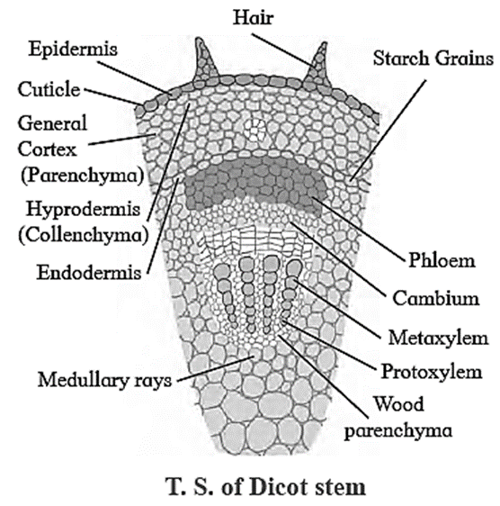
Anatomy of Dicot Stem (Sunflower) :

A transverse section of sunflower (dicot) stem shows the following structures:

Epidermis: It is a single, outermost layer with multicellular outgrowth called trichomes. A layer of cuticle is usually present towards the outer surface of epidermis.

Cortex: Cortex is situated below the epidermis and is usually differentiated into three regions namely, hypodermis, general cortex and endodermis.

  • Hypodermis: It is situated just below the epidermis and is made of 3-5 layers of collenchymatous cells. Intercellular spaces are absent.
  • General cortex: It is made up of several layers of large parenchymatous cells with intercellular spaces.
  • Endodermis: It is an innermost layer of cortex which is made up of barrel shaped cells. It is also called starch sheath, as it is rich in starch grain.

Stele: It is differentiated into pericycle, vascular bundles and pith.

  • Pericycle: It is the outermost layer of vascular system situated between the endodermis and vascular bundles. In sunflower, it is multi-layered and also called hard bast.
  • Vascular bundles: Vascular bundles are conjoint, collateral, open, and are arranged in a ring. Each One is composed of xylem, phloem and cambium. Xylem is endarch. A strip of cambium is present between xylem and phloem.
  • Pith: It is situated in the centre of the young stem and is made up of large-sized parenchymatous cells with conspicuous intercellular spaces.

[collapse]

Anatomy of Monocot Stem :

Anatomy of Monocot Stem (Maize Stem) :

A transverse section of maize (monocot) stem shows the following structures:

  • Epidermis: It is single layered and without trichomes.
  • Hypodermis: It is sclerenchymatous.
  • Ground tissue: It consists of thin walled parenchyma cells. It extends from hypodermis to the centre. It is not differentiated into cortex, endodermis, pericycle and pith.
  • Vascular bundles: Vascular bundles are numerous and are scattered in ground tissue. Each vascular bundle is surrounded by a sclerenchymatous bundle sheath. Vascular bundles are conjoint, collateral and closed (without cambium). Xylem is endarch and shows lysigenous cavity.
  • Pith: Pith is absent.

[collapse]

Dorsiventral leaf :

Anatomy of Leaf : Dorsiventral Leaf

Structure of dorsiventral leaf: The mesophyll tissue is differentiated into palisade and spongy parenchyma in a dorsiventral leaf. This type is very common in dicot leaf.

The different parts of this leaf are as follows:

Upper epidermis: It consists of a single layer of tightly packed rectangular, barrel shaped, parenchymatous cells which are devoid of chloroplast. A distinct layer of cuticle lies on the outside of the epidermis. Stomata are generally absent.

Mesophyll: Between upper and lower epidermis, there is chloroplast-containing photosynthetic tissue called mesophyll It is differentiated into Palisade parenchyma and Spongy parenchyma.

  • Palisade parenchyma: Palisade parenchyma is present below upper epidermis and consists of closely packed elongated cells. The cells contain abundant chloroplasts and help in photosynthesis.
  • Spongy parenchyma: Spongy parenchyma is present below palisade tissue and consists of loosely arranged irregularly shaped cells with intercellular spaces. The spongy parenchyma cells contain chloroplast and are in contact with the atmosphere through stomata.

Vascular system: It is made up of a number of vascular bundles of varying size depending upon the venation. Each one is ‘surrounded by a thin’ layer’ of parenchymatous cells called bundle sheath.

Vascular bundles are closed. Xylem lies towards upper epidermis and phloem towards lower epidermis.

Cambium is absent, hence there is no secondary growth in the leaf.

Lower epidermis: It consists of a single layer of compactly arranged rectangular, parenchymatous cells. A thin layer of cuticle is also present. The lower epidermis contains a large number of microscopic pores called stomata. There is an air-space called substomatal chamber at each stoma.

[collapse]

Isobilateral leaf :

Isobilateral Leaf :

The parts of isobilateral leaf are as follows.

Epidermis:

  • It is single layered, present on both sides of the leaf.
  • It consists of compactly arranged rectangular transparent parenchymatous cells.
  • Both the surfaces contain stomata.
  • Both the surfaces have a distinct layer of cuticle.

Mesophyll: Mesophyll is not differentiated into palisade and spongy tissue.

Vascular bundle: These are conjoint, collateral and closed.

[collapse]

Dorsiventral and Isobilateral leaf :

Compare between dorsiventral and isobilateral leaf :

Dorsiventral Leaf Isobilateral leaf
Dorsiventral Leaf is very common in dicotyledonous plants. Isobilateral leaf is very common in monocotyledonous plants.
In this mesophyll tissue is differentiated into palisade and spongy parenchyma. In this mesophyll tissue is not differentiated into palisade and spongy parenchyma.
The leaves are commonly horizontal in

orientation with distinct upper and lower surfaces. The upper surface which faces the sun is darker than the lower surface.

In this leaf both the surfaces are equally illuminated as both the surface can face the sun, and show similar structure. The two surfaces are equally green.
Stomata is absent on the upper surface of these leaves. Stomata is present on both the upper and lower surfaces

[collapse]

Rs 16

-Kitabcd Academy Offer-

Buy Notes(Rs.10)+ Solution(Rs.10) PDF of this chapter
Price : Rs.20 / Rs.16

Click on below button to buy PDF in offer (20% discount)

Useful Links

Main Page : – Maharashtra Board Class 11th-Biology  – All chapters notes, solutions, videos, test, pdf.

Previous Chapter : Chapter-7- Cell Division  – Online Notes

Next Chapter : Chapter-9-Morphology of Flowering PlantsOnline Notes

Leave a Reply

Write your suggestions, questions in comment box

Your email address will not be published. Required fields are marked *

We reply to valid query.