Notes-Part-1
|
Topics to be Learn : Part-1
|
Introduction :
Organisms move in a variety of ways. Movements range from protoplasm streaming to peristalsis to walking or running, among other things.
Movement and Locomotion :
Movements : Streaming of protoplasm, peristalsis are internal movements. Walking and running are external movements.
- Examples of internal movement: Contraction and relaxation of heart, inspiration and expiration, contraction of blood vessels, etc.
- Examples of external movement: Swimming; movement tongue, jaws, snout, tentacles; movement of ear pinna, etc.
Three muscle groups responsible for human movement :
- Smooth / non-striated / visceral / involuntary muscles
- Cardiac muscles
- Skeletal / striated / voluntary muscles.
- Involuntary muscles (smooth muscles and cardiac muscles) do not contract as per our will. Cardiac muscles show rhythmic contractions. Skeletal muscle is present in the diaphragm of the respiratory system.
Functions :
- Smooth muscles: They bring about involuntary movements like peristalsis in the alimentary canal constriction and dilation of blood vessels.
- Cardiac muscles: They bring about contraction and relaxation of the heart.
- Striated muscles: They control voluntary movements of limbs, head, trunk, eyes, etc.
- Skeletal muscles : are responsible for running and speaking.
Tendons : Skeletal muscles are attached to the bones by tendons and help in the movement of the parts of skeleton. Tendons are inelastic thick band of collagen fibers.
Locomotion : The change in locus of whole body of living organism from one place to another place is called locomotion.
Movement and locomotion is the combined action of bones, joints and skeletal muscles
The four basic types locomotory movements seen in animals are:
- Amoeboid movement: It is performed by pseudopodia. e.g. leucocytes.
- Ciliary movement: It is performed by cilia. e.g. ciliated epithelium. In Paramoecium, cilia help in passage of food through cytopharynx.
In trachea, ciliary movement removes the dust particles and foreign substance trapped. In female reproductive tract, passage of ova is facilitated by ciliary movement.
- Whirling movement: It is performed by flagella. e.g. sperms.
- Muscular movement: It is performed by muscles, with the help of bones and joints.
Q. All locomotions are movements but all movements are not locomotion. Justify
Ans: Locomotion occurs when body changes its position, however all movements may not result in locomotion. Thus, all locomotion are movements but all movements are not locomotion.
Location and structure of skeletal muscles :
Location: Major part of skeletal muscles which move the bones do not lie on the same bone but they are located on the bone atop. e.g. Biceps and triceps that move the forearm are located in the upper arm. -
Structure:
- At any joint, two types of bones are present i.e., stationary and movable.
- The end of the skeletal muscle to which the stationary bone is attached is called the origin whereas the opposite end attached to movable bone is called insertion.
- The middle thick part of the muscle is called as belly.
- All the fibres in a muscle do not extend from end to end and they are maximum in the middle part of the muscle. Thus, most of the large muscles are fusiform in shape.
Types of striated muscles :
On the basis of movements, striated muscles are of three types:
- Agonists : These are considered as the prime movers. They bring about the initial movement of a part e. g. biceps.
- Antagonists: They bring about the action opposite to that of prime movers. e.g. Triceps.
- Synergists: They assist the prime movers. e.g. Brachialis assist biceps.
Working of Skeletal Muscles :
Antagonistic muscles : The muscles that work in pairs and produce opposite action are known as antagonistic muscles. e.g. biceps and triceps of upper arm.
- The biceps (flexors) bring flexion (folding) and triceps (extensors) bring extension of the elbow joint.
- One member from a pair is capable of bending the joint by pulling of bones the other member is capable of straightening the same joint by pulling.
- In antagonistic pair of muscles, one member is stronger than the other. e.g. The biceps are stronger than the triceps.
Important antagonistic muscles:
- Flexor and extensor: Flexor muscle on contraction results into bending or flexion of joint. e.g. Biceps. Extensor muscle on contraction results in straightening or extension of a joint. e.g. Triceps.
- Abductor and adductor: Abductor muscle moves a body part away from the body axis. e. g. Deltoid muscle of shoulder moves the arm away from the body Adductor muscle moves a body part towards the body axis. e.g. Latissimus dorsi of shoulder moves the arm near the body.
- Pronator and supinator: Pronator turns the palm downwards and supinator turns the palm upward.
- Levator and depressor: Levator raises a body pan and the depressors lower the body part.
- Protractor and retractor : Protractor moves forward, whereas the retractor moves backward.
- Sphincters: Circular muscles present in the inner walls of anus, stomach, etc., for closure and opening
Structure of myosin and actin filaments :
Myosin filament:
- Each myosin filament is a polymerized protein, and one thick filament is made up of many meromyosins (monomeric proteins).
- A myosin molecule is made up of two heavy chains (heavy meromyosin/HMM) that coil around each other to form a double helix.
- A cross bridge is one end of each of these chains that is projected outwardly. This end folds to form the myosin head, a globular protein mass.
- Each head has two light chains, resulting in four light chains/light meromyosin/LMM.
- Myosin heads have ATPase activity. It can split ATP to generate energy.
- Myosin accounts for 55% of muscle proteins.

Actin filament: It is a complex type of contractile protein. It is made up of three components.
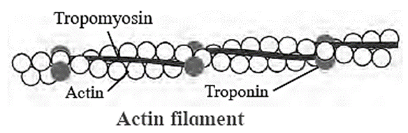
- F actin: This protein serves as the backbone of actin filaments. F actin consists of two helical strands. Each strand is made up of G actin molecules that have been polymerized. One ADP molecule is bound to one G actin molecule.
- Tropomyosin: The actin filament contains two additional protein strands that are tropomyosin polymers. Each strand is only loosely connected to a F actin. Tropomyosin physically covers the actin strand's active myosin-binding site when it is at rest.
- Troponin: It is a complex of three globular proteins, is attached approx. 2/3rd distance along each tropomyosin molecule. It has affinity for actin, tropomyosin and calcium ions. The troponin complex is believed to attach the tropomyosin to the actin. The strong affinity of troponin for calcium ions is believed to initiate the contraction process.
Mechanism of muscle contraction :
Sliding filament theory: It was put forth by H.E Huxley and A.F Huxley. It is also known as ‘Walk along theory’ or Ratchet theory.
- The sliding filament theory proposes that the interaction of actin and myosin filaments is the fundamental cause of muscle contraction. Myosin filaments are interdigitated with actin filaments.
- A cross bridge connects the head of myosin to the actin backbone, forming a hinge joint. Myosin head cannot tilt forward or backward from this joint. This movement is an active process because it makes use of ATP.
- ATPase activity is present in the myosin head. It can obtain energy from the breakdown of an ATP molecule. This energy can be used to move the myosin head.
- During contraction, the myosin head binds to the active site of actin filaments and pulls them inward, allowing the actin filaments to slide over the myosinfilaments. This causes muscle fibres to contract.

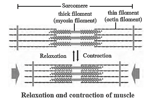
Sarcomere : Sarcomere is the functional unit of myofibril. It has specific arrangement of actin and myosin filaments. The components of sarcomere are organized into variety of bands and zones.
The structure of sarcomere:
- ‘A’ band — dark bands present at the centre of sarcomere and contain myosin as well as actin.
- ‘H’ zone or Hensen s zone — light area present at the centre of ‘A’ band
- ‘M’ line — present at the centre of ‘H’ zone -
- ‘I’ band — light bands present on the either side of ‘A’ band containing only actin
- Z line — adjacent ‘I’ bands are separated by ‘Z’ line.
Know This : The fundamental characteristic of the muscle is contraction. Therefore, muscle can only pull and not push the bone.
Physiology of muscle Contraction :
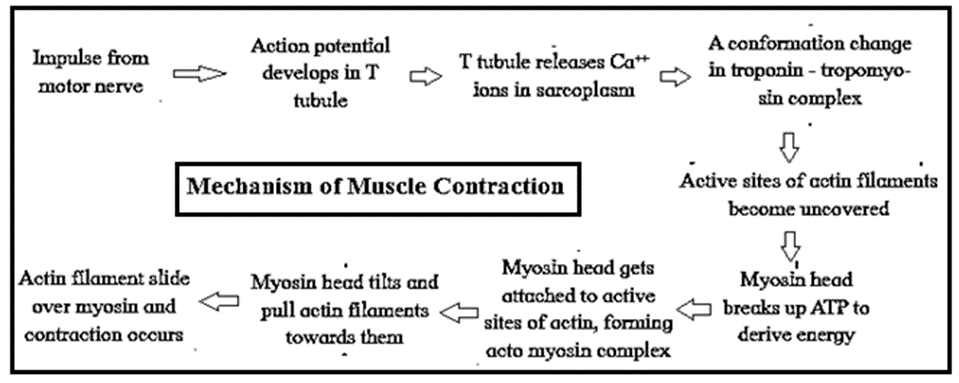
- When the muscles are relaxed, tropomyosin and troponin complex remain on the active sites. As a result, myosin is unable to interact with the active site of actin, preventing contraction.
- When an impulse (action potential) enters the muscle via the motor end plate, it spreads throughout the myofibril's sarcolemma.
- The sarcoplasmic reticulum's transverse tubules release a large number of Ca++ ions into the sarcoplasm. These calcium ions interact with troponin molecules, inactivating the troponin-tropomyosin complex. Tropomyosin's structure changes as a result of this.
- As a result, tropomyosin becomes detached from the actin (F actin) filament's active site. As a result, the active site of actin is revealed.
- To obtain energy, the myosin head cleaves ATP and attaches to the uncovered active site of actin.
- As a result, an acto-myosin complex is formed.
- Myosin heads are now tilted backwards, drawing the attached actin filament inward. This causes the muscle fibres to contract.
| Know This :
T tubules : T tubules or the transverse tubules are invaginations of the sarcolemma penetrating into the myocyte interior, forming a highly branched and interconnected network that makes junctions with the sarcoplasmic reticulum. These tubules are selectively enriched with specific ion channels and proteins crucial in the development of calcium transients necessary in excitation-contraction coupling, thereby facilitating a fast-synchronous contraction of the entire cell volume. They are unique to striated muscle cells. |
Relaxation of muscle fibres :
Muscle relaxation:
Muscle contraction and relaxation are active processes as during both the processes energy is utilized by hydrolysis of ATP into ADP and inorganic phosphate by the enzyme ATPase.
Muscle relaxation process : During relaxation, all the events occur in reverse direction as that of muscle contraction.
- When the stimulation is terminated, the actomyosin complex is broken down and myosin head gets detached from actin filaments. This process utilizes ATP.
- Also, the Ca++ ions are pumped back into the sarcoplasmic reticulum. This process too is an energy dependent process and utilizes ATP.
- As a result, the troponin-tropomyosin complex is restored again which covers the active sites of actin filament, due to disappearance of the Ca++ ions.
- The interaction between actin and myosin ceases and the actin filaments return back to their original position.
- This results in muscle relaxation.
Calcium ions : Calcium ions play a major role in contraction and relaxation of muscles. Calcium ions are released from the sarcoplasm during muscle contraction and stored in sarcoplasm during muscle relaxation.
Properties of Muscles on Electrical Stimulation:
(i) Single muscle twitch : A muscle contraction initiated by a single brief-stimulation is called a single muscle twitch.
It occurs in 3 stages :
- a latent period of no contraction,
- a contraction period
- a relaxation period.
(ii) Summation : If the muscle is stimulated before the end of the twitch, it generates greater tension i.e., summation or addition of effect takes place. Repeated stimuli will produce increasing strength of contraction (stair case phenomenon).
(iii) Tetanus : If stimulation is very rapid and frequent the muscle does not have time to relax. It remains in a state of contraction called tetanus.
(iv) Refractory period : Immediately after one stimulus, the muscle fibre cannot respond to another stimulus. This resting or refractory period is about 0.02 seconds.
(v) Threshold stimulus : For a muscle fibre to contract, a certain minimum strength or intensity of stimulus is required. This is called threshold stimulus.
(vi) All or none principle :
- A stimulus below the threshold will not cause contraction. A threshold stimulus causes contraction.
- This contraction produces the greatest amount of force.
- A higher stimulus will not increase the force of contraction, implying that a muscle fibre will either contract completely or not at all.
- This is the 'all or nothing' principle. This law applies to all muscle and nerve fibres.
(vii) Oxygen debt :
- During strenuous exercise, the oxygen supply to the muscle quickly becomes insufficient to sustain oxidative phosphorylation of the respiratory substrate.
- Muscles contract anaerobically at this stage and accumulate lactic acid produced by anaerobic glycolysis.
- Lactic acid is toxic and produces less ATP. It causes fatigue, pain, and muscle cramps.
- The oxygen consumption of the muscle during recovery far exceeds that of the resting state.
- The extra oxygen consumed during recovery is referred to as muscle oxygen debt.
| Know This :
Rigor Mortis : Usually, some hours after the death of an individual, its muscles are stiffened. This muscular stiffening, after death is rigor mortis. It helps in fixation of hours of death after a murder. After death, the fresh supply of ATP to muscles becomes impossible. Therefore once the local store of ATP is finished, the detachment of myosin from actin cannot take place. This results in permanent state of contraction of the muscle. Oxygen debt : It is used in oxidizing the accumulated lactic acid aerobically and in restoring the depleted creatine phosphate and ATP. |
Skeletal System :
The components of skeletal system are bones, tendons, ligaments and joint.
Endoskeleton and Exoskeleton :
- When supportive structures are present inside the body, they form the endoskeleton, When supportive structures are present on the body's outer surface, they form the exoskeleton.
- Bones and cartilage form major endoskeletal components
Exoskeleton :
Exoskeleton components : Exoskeleton components change from lower to higher group of animals. These include chitinous structures, nails, horns, hooves, scales, hair, shell, plates, fur, muscular foot, tube feet, etc.
- Nails, hooves, scales, plates, muscular foot and tube feet help in movement and locomotion.
- Scales and plates in reptiles like snakes provide grip to move on rough edgy surfaces.
- Fishes have dermal scales (bony scales), whereas reptiles like snakes have epidermal scales or scutes (horny, tough extensions of outer layer of skin i.e., stratum corneum).
Exoskeletal structures:
- Exoskeleton provide support, help in movement and also provides protection from predators.
- The exoskeletal structures vary from organism to organism. Echinoderms have tube feet for locomotion whereas molluscs (e.g. Chiton) have muscular foot for movement and locomotion.
Bones :
Type of bones present in our body :.
Long bones, short bones, flat bones, irregular bones and sesamoid bones.
Functions of bones :
- Bones form the framework of our body and thus provide shape to the body
- They protect vital organs thus help in the smooth functioning of body.
- The joints between the bones help in movement and locomotion.
- They provide firm surface for attachment of muscles.
- They are reservoirs of calcium and form important site for hemopoiesis.
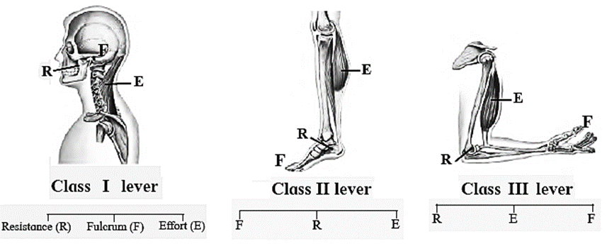
The lever system in human body:
In the human body, the joints act as fulcrums and the bones act as levers.
The respective muscles generate force required to move the load i.e. the bone along with the overlying tissues and anything else that has to be moved along with it,
Types of lever found in human body :
The three types of lever are as follows:
Class I lever: The joint between the first vertebra and occipital condyle of skull is an example of Class I lever. The force is directed towards the joints (fulcrum); contraction of back muscle provides force while the part of head that is raised acts as resistance.
Class II lever: Human body raised on toes is an example of Class II lever. Toe acts as fulcrum, contracting calf muscles provide the force while raised body acts as resistance.
Class III lever: Flexion of forearm at elbow exhibit lever of class III. Elbow joint acts as fulcrum and radius, ulna provides resistance. Contracting bicep muscles provides force for the movement.
We reply to valid query.