Solution-Class-11-Science-Biology-Chapter-11-Study of Animal Type-Cockroach-Maharashtra Board

-Study of Animal Type-Cockroach

Maharashtra Board-Class-11-Science-Biology-Chapter-11

Solution

Question 1.

Choose correct option

(A) Chemical nature of chitin is .............

(a) protein.

(b) carbohydrate.

(c) lipid.

(d) glycoprotein.

Answer :

(b) carbohydrate.

(B)  Cockroach has ................ type of mouthparts.

(a)  sponging

(b)  chewing and biting

(c)  piercing and Sucking

(d)  lapping

Answer :

(b)  chewing and biting

(C)  Spiracle is a part of ................ system of cockroach.

(a)  circulatory

(b)  respiration

(c)  reproductive

(d)  nervous

Answer :

(b)  respiration

(D)  ................... is a part of digestive system.

(a)  Trachea

(b)  Hypopharynx

(c)  Haemocyte

(d)  Seminal vesicle

Answer :

(b)  Hypopharynx

(E)  ..................... is also called as brain of cockroach.

(a)  Supra-oesophageal ganglion

(b)  Sub-oesophageal ganglion

(c)  Hypo-cerebral ganglion

(d)  Thoracic ganglion

Answer :

(a)  Supra-oesophageal ganglion

Question 2.  

Answer the following questions

(A) Describe the digestive system of cockroach.

Answer :

Digestive System of Cockroach :

Digestive system of cockroach consists of mouthparts, alimentary canal and salivary glands.

Mouth parts : Pre-oral cavity present in front of the mouth receives food. It is bounded by chewing and biting type of mouth parts. These are movable, segmented appendages that help in ingestion of food.

The mouthparts of cockroach comprises of:

Labrum: It forms the upper lip. It is a single flap-like movable part which covers the mouth from upper side. It forms an anterior wall of pre-oral cavity.

  • Function : It is useful in holding the food during feeding.

Mandibles: These are two dark, hard, chitinous structures with serrated median margins. They are true jaws present on either side, behind the labrum.

  • Function: They perform co-ordinated side-wise movements with the help of adductor and abductor muscles to cut and crush the food.

Maxillae: These are the accesssory jaws. They are also called as first pair of maxillae. These are situated on the either side of mouth behind the mandibles. Each maxilla consists of sclerites like cardo, stipes, galea, lacinia and maxillary palps.

  • Functions: Maxillae hold food, help mandibles for mastication. They are also used for cleaning the antennae and front legs. Maxillary palps act as tactile organs.

Labium: It forms the lower lip. Labium is also known as second maxilla which covers the pre-oral cavity from the ventral side. It is firmly attached to the posterior part of head. It has three jointed labial palps which are sensory in function.

  • Function: It is useful in pushing the chewed food in the pre—oral cavity. It prevents the loss of food falling from the mandibles, while chewing.

Hypopharynx : Hypopharynx / tongue / lingua is a somewhat cylindrical single structure, located in front of the labium and between first maxillae. A salivary duct opens at the base of Hypopharynx. Hypopharynx bears comb-like plates called super-lingua on either side.

  • Function: It is useful in the process of feeding and mixing saliva with food.

Alimentary canal: It is long (6 - 7cm) tube of different diameters and two openings

Cockroach does not have the buccal cavity thus, the alimentary canal begins from pre-oral cavity

The alimentary canal is divisible into three parts: foregut, midgut and hindgut.

(i) Foregut or stomodaeum : It consists of pharynx, oesophagus, crop and gizzard.

  • Pharynx: It is very short, narrow but muscular tube that opens into oesophagus. It containstaste sensillae. Function: Conduction of food into the oesophagus
  • Oesophagus: It is slightly long and narrow tube which opens into crop.
  • Crop: Crop is a large, pear shaped and sac-like organ. Function: It temporarily stores the food and then sends it to gizzard.
  • Gizzard: Gizzard or proventriculus is a small spherical organ. It is provided internally with a circlet of six chitinous teeth and backwardly directed bristles. The foregut ends with gizzard. Function: The chitinous teeth present ingizzard are responsible for crushing the foodand the bristles help to filter the food.

(ii) Midgut or mesenteron: It consists of stomach and hepatic caeca.

  • Ventriculus or stomach: It is straight, short and narrow. Stomach is _lined by glandular epithelium which secretes digestive enzymes. Function: It is mainly responsible for digestion and absorption.
  • Hepatic caeca: These are thin, transparent, short, blind (closed) and hollow tubules. Function: They secrete digestive enzymes.

(iii) Hindgut or proctodaeum : It consists of ileum, colon and rectum.

  • Ileum: It is short and narrow part of hindgut. Malpighian tubules open in the anterior lumen of ileum, near the junction of midgut and hindgut. Posterior region of ileum contains sphincter. Ileum directs the nitrogenous wastes and undigested food towards colon.
  • Colon: It is a longer and wider part of the hindgut. It directs waste material towards the rectum. It reabsorbs water from wastes as per the need.
  • Rectum: It is oval or spindle-shaped, terminal part of the hindgut. It contains six rectal pads along the internal surface for absorption of water. Rectum open into anus. Anus is present onthe ventral side of the 10th segment. It is the last or posterior opening of the digestive system. The undigested food is released out of the body through anus.

Salivary glands:

  • Cockroach has a pair of salivary glands which secrete saliva.
  • Each salivary gland has two glandular lobes and a receptacle or reservoir.
  • The glandular lobes consists of several irregular-shaped white coloured lobules which secrete saliva.
  • Each gland has a salivary duct. Both the ducts unite to form a common salivary duct.
  • Receptacle of each salivary gland is thin-walled, elongated, sac-like structure. Each receptacle has a duct. These ducts unite to form common reservoir duct.
  • Common salivary duct and common reservoir duct unite together to form a common efferent salivary duct. The efferent salivary duct opens at the base of tongue or hypopharynx.

(B) Give an account on tracheal system of cockroach?

Answer :

Respiratory System or Tracheal System :

Cockroaches have an internal respiratory system of air tubes called the tracheal system, which brings air into the body and makes contact with every pan. It enables direct gas exchange between air and tissues without the use of blood.

These air tubes of internal respiratory system begin at the opening on body surface called spiracles.

  • Spiracles: They are paired respiratory openings. Spiracles are present on the ventro-lateral side of the body, of thoracic and eight pairs of abdominal spiracles. The in pleural membrane. Cockroaches have two pairs spiracles open into a series of air sacs from which arise the tubes called trachea.
  • Trachea: The trachea form a definite pattern of branching tubes arranged transversely as well as longitudinally. They are about 1 mm thick and have spiral or annular thickening of chitin. The inner lining chitin prevents the trachea from collapsing. Each trachea further branches into smaller tubes called tracheoles.

Tracheoles : These are fine intracellular tubes that penetrate deep into tissues. They are thin and not lined by chitin. They end blindly in the cells. Each tracheole at the blind end is filled with a watery fluid through which exchange of gases takes place. The content of the fluid keeps changing. At high muscular activity, part of fluid part is drawn into the tissues to enable more and rapid oxygen intake.

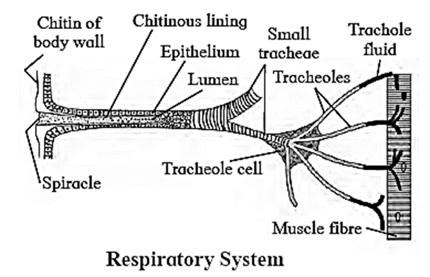
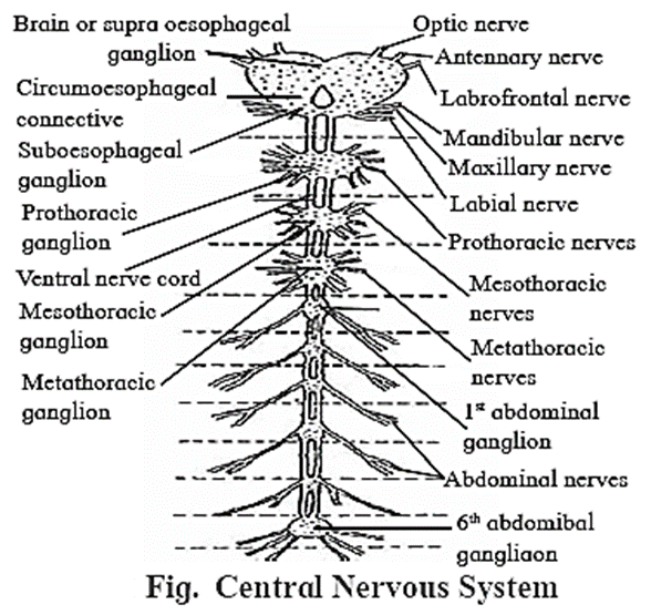
(C) Describe nervous system of cockroach.

Answer :

Nervous System :

Nervous system of cockroach is ventral, solid and ganglionated. It consists of central nervous system (CNS), peripheral nervous system (PNS) and autonomous nervous system (ANS).

  • The nervous system of cockroach is spread throughout the body.
  • The head of cockroach holds a bit of nervous system while the rest is situated along the ventral part of its body.

Central nervous system (CNS): Central nervous system consists of (1) nerve ring and (2) ventral nerve cord.

(1) Nerve ring consists of:

  • a pair of supra-oesophageal ganglia
  • a pair of circum-oesophageal connectives
  • a pair of sub-oesophageal ganglia

(i) Supra-oesophageal ganglia or cerebral ganglia : A pair of supra-oesophageal ganglia is collectively known as the brain. Brain is present in head, above the oesophagus and between antennal bases. Each supra-oesophageal ganglion is formed by the fusion of three small ganglia - protocerebrum, deutocerebmm and tritocerebrum.

(ii) Circum-oesophageal connectives: Supra-oesophageal ganglia are connected to sub-oesophageal ganglion by a pair of lateral nerves called as circum-oesophageal connectives. Connectives arise from supra-oesophagial ganglia.

(iii) Sub-oesophageal ganglia: It is a bilobed and present below the 0esophagus, in head. It is also formed by the fusion of three pairs of ganglia.

(2) Ventral nerve cord :

  • It arises from the sub-oesophageal ganglion. It is present along mid-ventral position, in perineural sinus.
  • It is double ventral nerve cord and consists of nine segmental, paired ganglia. .
  • First three pairs of segmental ganglia are large and known as thoracic ganglia. The other six pairs of segmental ganglia are in abdomen (abdominal ganglia).
  • 6th abdominal ganglion is the largest and it is present in 7th abdominal segment.
  • There is no ganglion in 6th segment.

Peripheral nervous system (PNS):

  • The peripheral nervous system comprises of nerves that arise from various ganglia of CNS.
  • Six pairs of nerves arise from the supra-oesophageal ganglia.They supply to the eyes, antenna and labrum.
  • Nerves arising from the sub-oesophageal ganglion supply to the mandibles, maxillae and labium
  • Nerves arising from the thoracic ganglia supply to the wings, legs and intemal thoracic organs.
  • Nerves from abdominal ganglia go to the abdominal organs of respective abdominal segments.

Autonomic nervous system (ANS): It consists of four ganglia and a retro cerebral complex.

The ganglia are as follows:

  • Frontal ganglion: It is present above the pharynx and in front of brain.
  • Hypocerebral ganglion: It is present on the anterior region of oesophagus.
  • Ingluvial ganglion: It is present on crop. It is also called as visceral ganglion.
  • Ventricular ganglion: It is present on gizzard.

(D) With help of neat labelled diagram, describe female reproductive system of cockroach.

Answer :

Female reproductive system :

  • Female reproductive system consists of primary and secondary reproductive organs.
  • Ovaries are primary reproductive organs. They are paired and lie lateral in position in 2nd - 6th abdominal segments. Each ovary is formed of a group of eight ovarian tubules or ovarioles, containing a chain of developing ova.
  • All ovarioles of an ovary open in lateral oviduct of respective side.
  • Both the lateral oviducts unite to form a common oviduct or vagina.
  • Common oviduct or vagina opens into the Bursa copulatrix (genital chamber), the female organ of copulation.

In females, the 7th sternum is boat shaped and together with 8th and 9th sternum forms a brood or genital pouch whose anterior pan contains gonopore, spennathecal pores and collateral glands.

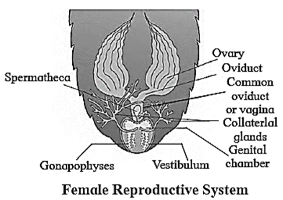
(E) With help of neat labelled diagram, describe the digestive system of cockroach.

Answer :

Digestive System of Cockroach :

Digestive system of cockroach consists of mouthparts, alimentary canal and salivary glands.

Mouth parts : Pre-oral cavity present in front of the mouth receives food. It is bounded by chewing and biting type of mouth parts. These are movable, segmented appendages that help in ingestion of food.

Alimentary canal: It is long (6 - 7cm) tube of different diameters and two openings

Cockroach does not have the buccal cavity thus, the alimentary canal begins from pre-oral cavity. The alimentary canal is divisible into three parts: foregut, midgut and hindgut.

Salivary glands:

  • Cockroach has a pair of salivary glands which secrete saliva.
  • Each salivary gland has two glandular lobes and a receptacle or reservoir.
  • The glandular lobes consists of several irregular-shaped white coloured lobules which secrete saliva.
  • Each gland has a salivary duct. Both the ducts unite to form a common salivary duct.
  • Receptacle of each salivary gland is thin-walled, elongated, sac-like structure. Each receptacle has a duct. These ducts unite to form common reservoir duct.
  • Common salivary duct and common reservoir duct unite together to form a common efferent salivary duct. The efferent salivary duct opens at the base of tongue or hypopharynx.

[Also refer Q.2(A)]

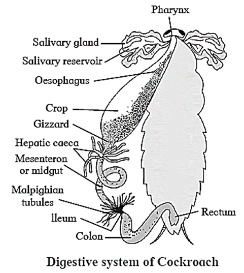
(F) A student observed that the cockroaches are killed for dissection by simply putting them in soap water. He inquired whether soap is so poisonous. Teacher said it is due to its peculiar respiratory system. How?

Answer :

When cockroaches are immersed in soap solution, the solution enters their bodies through small respiratory openings known as spiracles. As a result, cockroaches may be unable to breathe and may suffocate.

(G) Describe the circulatory system of cockroach.

Answer :

Circulatory System or Blood Vascular System :

Cockroach has open circulatory system. It consists of colourless blood (haemolymph), a dorsal blood vessel (heart and dorsal aorta) and haemocoel.

Haemolymph: Haemolymph is colourless as it is without any pigment. It consists of plasma and seven types of blood cells/haemocytes. Plasma consists of water with some dissolved organic and inorganic solutes. It is rich in nutrients and nitrogenous wastes like uric acid.

Haemocoel: The body cavity of cockroach (haemocoel) can be divided into three sinuses due to two diaphragms i.e. dorsal and ventral diaphragm. These diaphragms are thin, fibromuscular septa (sing.septum). It remains attached to terga along lateral sides at intermittent points.

(i) Dorsal diaphragm : It has 12 pairs (10 abdominal and 2 thoracic) of fan-like alary muscles. Alary muscles are triangular with pointed end attached to terga at lateral side and broad end lies between the heart and dorsal diaphragm.

(ii) Ventral diaphragm: It is flat and present just above the ventral nerve cord. Laterally, it is attached to stema at intermittent points.

(iii) Sinuses: The coelom of cockroach is divided into three sinuses — pericardial sinus, perivisceral sinus and perineural sinus.

  • Pericardial sinus : It is dorsal, very small and contains dorsal vessel. _
  • Perivisceral sinus : It is middle and largest sinus. It contains fat bodies and almost all major visceral organs of alimentary canal and reproductive system.
  • Perineural sinus : It is ventral, small and contains ventral nerve cord. It is continuous into legs. All the three sinuses communicate with each other through the pores present between two successive points of attachments of diaphragms.

Dorsal blood vessel : This is present in pericardial sinus, just below the tergum. It is divisible into posterior heart and anterior aorta (dorsal aorta/cephalic vessel). _ .

  • Heart : It is about 2.5 cm long, narrow, muscular tube that is open anteriorly and closed posteriorly. It starts from 9th  abdominal segment and extends anteriorly upto 1st  thoracic segment. Heart of cockroach is 13 chambered, out of which 10 chambers are in abdominal region and three are in thoracic region. Each chamber has a pair of vertical slit-like incurrent aperture or opening called ostium (plural: ostia). Ostia are present along lateral side in the posterior region of first 12 chambers;Each ostium has lip-like valves that allow the flow of blood from sinus to heart only.

Anterior aorta : Heart is continued by a short, thin-walled vessel called dorsal aorta. It lies in head region and opens in haemocoel.

Question 3.

Answer the following questions

(A) How will you identify male or female cockroach?

Answer :

Male cockroach Female cockroach
Abdomen is relatively long and narrow Abdomen is shod and broad
7th tergum covers 8th tergum 7th tergum covers 8th  and 9th terga
Antennae are longer in size Antennae are shorter in size
Anal styles are present Anal styles are absent
Brood pouch is absent Brood pouch is present
All 9 stema visible Only 7 sterna visible

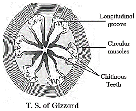
(B) Write a note on : Gizzard of cockroach.

Answer :

Gizzard: Gizzard or proventriculus is a small spherical organ. It is provided internally with a circlet of six chitinous teeth and backwardly directed bristles. The foregut ends with gizzard.

Function: The chitinous teeth present in gizzard are responsible for crushing the food and the bristles help to filter the food.

(C) Give the systematic position of cockroach.

Answer :

Systematic Position :

Classification Reason
Kingdom : Animalia Cell wall absent, heterotrophic nutrition.)
Phylym : Arthropoda Jointed appendages are present, segmented body, chitinous exoskeleton.
Class : Insecta Two pairs of wings and three pairs of walking legs are present.
Genus : Periplaneta Nocturnal, straight wings.
Species : americana Origin is in Continent of America

(D) What would have happened if cockroach did not had gizzard?

Answer :

  • The gizzard in cockroach is a spherical organ which has chitinous teeth and bristles.
  • The chitinous teeth present in gizzard are responsible for crushing the food and the bristles help to filter the food.
  • If the cockroach did not have gizzard, the food .will not be crushed into small particles and unfiltered food will enter the hindgut.

(E) What is the functional difference between eyes of cockroach and human being?

Answer :

  • Cockroach eyes have several ommatidia that form an image and allow them to detect even the smallest movement of their predator, whereas human eyes can only look in one direction at a time.
  • They have binocular vision, which gives them a better sense of depth and a three-dimensional image of their surroundings.

(F) What is the functional difference between respiratory systems of cockroach and human being?

Answer :

  • The functional difference between the cockroach and human respiratory systems is that the cockroach respiratory system allows the exchange of gases directly between the air and tissues without the need for transportation by blood, whereas the human respiratory system requires blood to transport gases to the body tissues.
  • Thus, the circulatory system in cockroaches plays no role in the respiratory process, whereas the circulatory system in humans plays an important role in the respiratory process.

Question 4.

Explain the following in short

(A) What are anal cerci?

Answer :

Anal cerci are a pair of appendages at the end of the abdomen that arise from the 10th segment of the body of cockroach.

(B) What is the ganglion?

Answer :

Ganglion is a group of nerve cell bodies.

(C) What is the role of hypopharynx?

Answer :

Hypopharynx : Hypopharynx / tongue / lingua is a somewhat cylindrical single structure, located in front of the labium and between first maxillae. A salivary duct opens at the base of Hypopharynx. Hypopharynx bears comb-like plates called super-lingua on either side.

Hypopharynx is useful in the process of feeding and mixing saliva with food.

(D) What is mesenteron?

Answer :

Midgut or mesenteron: It consists of stomach and hepatic caeca.

  • Ventriculus or stomach: It is straight, short and narrow. Stomach is lined by glandular epithelium which secretes digestive enzymes. It is mainly responsible for digestion and absorption.
  • Hepatic caeca: These are thin, transparent, short, blind (closed) and hollow tubules. They secrete digestive enzymes.

(E) Location of turgum.

Answer :

  • Tergum is located in abdomen of cockroach
  • Tergum is jointed to the stemum laterally by a soft cuticle called pleura

(F) What is ootheca?

Answer :

Ootheca :

  • The secretion of collaterial glands forms a capsule around them is called as ootheca or egg case.
  • It is about 8 mm long and ranges from dark reddish to blackish brown.
  • Ootheca contains 14 to 16 fertilized eggs in two rows.
  • They are dropped or glued to a suitable surface, like a crack or crevice of good humidity near the food source.
  • A female cockroach on an average, produces 9 to 10 oothecae during its lifespan.


(G) How many chambers are present in heart of cockroach?

Answer :

13 chambers are present in heart of a cockroach, out of which 10 chambers are in abdominal region and three are in thoracic region.

Rs 16

-Kitabcd Academy Offer-

Buy Notes(Rs.10)+ Solution(Rs.10) PDF of this chapter
Price : Rs.20 / Rs.16

Click on below button to buy PDF in offer (20% discount)

Useful Links

Main Page : – Maharashtra Board Class 11th-Biology  – All chapters notes, solutions, videos, test, pdf.

Previous Chapter : Chapter-10-  Animal Tissues – Online Solution

Next Chapter : Chapter-12-Photosynthesis  – Online Solution

Leave a Reply

Write your suggestions, questions in comment box

Your email address will not be published. Required fields are marked *

We reply to valid query.