Notes-Part-1-Class-12-Biology-Chapter-8-Respiration and Circulation-Maharashtra Board

Respiration and Circulation

Maharashtra Board-Class-12th-Biology-Chapter-8

Notes-Part-1

Topics to be Learn : 

  • Respiration
  • Organs of respiratory exchange
  • Human respiratory system
  • Mechanism of respiration
  • Regulation of breathing
  • Modified respiratory movements
  • Common disorders of respiratory system
  • Transportation in living organisms
  • Circulation in animals
  • Circulatory system in human
  • Red blood corpuseles / erythrocytes
  • White blood corpuseles / leucocytes
  • Thrombocytes / platelets

Introduction : The energy that is stored in the body in the form of complex organic compounds (potential energy) is however not usable by the organisms unless it is converted into usable form. This conversion is achieved through the process of respiration.

Respiration : Respiration is a biochemical process of oxidation of organic compounds in an orderly manner for the liberation of chemical energy in the form of ATP

Example : C6H12O6 + 6O2 → 6CO2 + 6H2O + 38 ATP  (In this, the process of gaseous exchange takes place between the organism and the environment.)

Organs of respiratory exchange :

Respiratory surface should possess the following features for efficient gaseous exchange.

  • A large surface area.
  • Thin, highly vascular and permeable to allow exchange of gases. .
  • Moist surfaces.

Gaseous exchange in plants : A terrestrial plant has stomata on leaves and young stems and lenticels on the stem surface for exchange of gases.

Respiration in Animals : In animals, depending upon the complexity of organization and the surrounding medium, respiratory organs have become specialized and are usually associated with a transport system.

Respiratory organs in different organisms :

Respiratory organs in different organisms :

1) Aquatic organisms :

Organisms Respiratory surface/ organ
Protists, Sponges and Coelenterates Plasma membrane
Limulus (Arthropod) Book gills
Amphibian tadpoles of frog, salamanders and newts External gills
Fish Internal gills
Flatworms like Planaria, Annelids (earthworm, nereis, leech), amphibians (frog) Plasma membrane, general body surface (moist skin)

 2) Aquatic organisms :

Organisms Respiratory surface/ organ
Insects Tracheal tubes and spiracles
Arachnids like spiders and scorpions Book lungs
Reptiles, Birds and Mammals Lungs

 3) Underwater organisms : Turtles - Respiratory surface/ organ : cloaca

  • Only at the time of diving or when underwater, turtles perform cloacal respiration. There are a pair of accessory air bladders connected to the cloaca which can absorb oxygen from the water.

[collapse]

Human respiratory system : Human respiratory system consists of nostrils, nasal chambers, pharynx, larynx, trachea, bronchi, bronchioles, lungs, aided by diaphragm and intercostal muscles.

1) Nostrils and nasal chambers :

1) Nostrils and nasal chambers : The nostrils are external openings of the nose.

  • Oxygen rich air is taken into the body through the nostrils or external nares. Carbon dioxide and water vapour are released out of the body through the nostrils.
  • The internal nares open into the pharynx. The space between the external and internal nares is known as nasal chamber. The nasal chamber is lined internally by mucous membrane and ciliated epithelium.
  • The nasal chamber is divided into right and left parts by a cartilage called mesethmoid.

Every nasal chamber is further divided into three regions : (i) Vestibule (ii) Respiratory part (iii) Sensory part.

  • Vestibule : The anteriormost part of the nasal chamber is vestibule. Hair present in this chamber prevent the dust particles from going inside.
  • Respiratory part : It is the part which is richly supplied by capillaries. Air is made warm and moist in this region.
  • Sensory part : The sensory epithelium lines this region. It is concerned with the detection of smell.

[collapse]

2) Pharynx : The pharynx is a short, vertical tube about 12 cm in length.

  • The respiratory and food passages cross each other in the pharynx,
  • The upper part of the pharynx is known as naso-pharynx. It conducts the air.
  • The lower part is called laryngo-pharynx or oro-pharynx. It conducts food to the oesophagus.
  • The tonsils are present in the pharynx. They are made of lymphatic tissue. They kill the bacteria that are trapped in mucus.

3) Larynx :

3) Larynx: The larynx produces sound.

  • In males, it increases in size at puberty. This is termed as Adam’s apple.
  • From the pharynx air enters the larynx. The opening through which it enters is called glottis.
  • The glottis has a guarding flap called epiglottis.
  • The epiglottis prevents the entry of food particles into the trachea.
  • The vocal cords are seen along the side of the glottis. They are made of elastic tissue. They produce sound.
  • Passage of air between the vocal cords and modulations created by tongue, teeth, lips and nasal cavity produce voice.

[collapse]

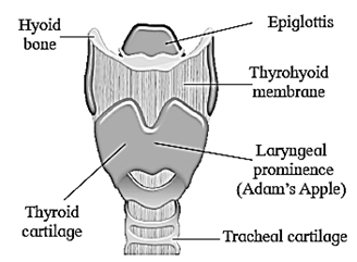
4) Trachea : The trachea or windpipe is about 10-12 cm long and 2.5 cm wide.

  • It is situated in front of the oesophagus and runs downwards in the thorax.
  • Fibrous muscular tissue supported by ‘C’ shaped cartilages form the walls of the trachea.
  • 16 to 20 cartilage rings are present in the trachea.
  • The trachea is lined internally by ciliated epithelium and mucous glands.
  • Mucous and ciliary action remove the dust particles and push them upwards to the larynx. These particles are then gulped and taken into the oesophagus. Instant coughing can remove foreign particles that enter the trachea.

5) Bronchi and bronchioles : The trachea divides into two bronchi (singular-bronchus) at its distal end behind the sternum.

  • The bronchus has complete ring of cartilage for support.
  • The bronchi enter the lungs on either side.
  • After entering the lungs each bronchus divides into secondary and tertiary bronchi. The tertiary bronchi divide further to form bronchioles.
  • The bronchioles are minute and are without the cartilage rings in their walls.
  • Each bronchiole ends into a bunch of alveoli which appear like a bunch of grapes. Each alveolus is balloon shaped.
  • Many alveoli make the lung spongy and elastic.

6) Lungs :

6) Lungs :

  • The lungs are principal respiratory organs located in the thoracic cavity.
  • They are pinkish, soft, hollow, paired, elastic and distensible organs.
  • The lung is enclosed in a pleural sac.
  • The pleural sac has two membranes—an outer parietal and an inner visceral which enclose the pleural cavity.
  • The pleural fluid which is present in the pleural cavity lubricates and prevents friction when pleural membranes slide on each other.
  • The lungs are richly supplied with blood capillaries and hence are highly vascular organs.
  • The left lung has two lobes while the right lung has three lobes.
  • Each lobe has many bronchioles and alveolar sacs.

[collapse]

7) Alveoli :

  • The alveolar sacs are spherical and thin walled and contain about 20 alveoli.
  • The alveoli are covered by a network of capillaries from pulmonary artery and pulmonary vein.
  • Each alveolus has thin and elastic wall. It is about 0.1 mm in diameter.
  • The alveolar wall is 0.0001 mm thick and is made of simple, non-ciliated, squamous epithelium. It has collagen and elastin fibres.
  • Every lung has about 700 million alveoli which increase the surface area for the exchange of gases.
  • The outermost covering of the lungs which made of smooth is known as visceral pleura is muscle fibres.
  • The lobule in the lung consists of alveolar ducts, alveolar sacs and alveoli. In alveoli gaseous exchange takes place.

8) Diaphragm : It is a muscular septum that separates the thoracic and abdominal cavity. It is dome shaped and on contraction it becomes flattened.

Mechanism of respiration :

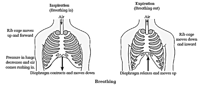
Mechanism of respiration :

The process of respiration includes breathing, external respiration, internal respiration and cellular respiration.

Breathing : Breathing is the process by which the air comes in and goes out of the lungs.

  • The rate of gaseous exchange is speeded up by breathing.
  • Breathing is a part of respiration and the terms breathing and respiration are not synonymous.
  • Inspiration and expiration together make breathing.

(i) Inspiration: Inspiration is an active process brought about by ribs, intercostal muscles, sternum and diaphragm.

  • The intercostal muscles contract and pull the ribs outwards. This increases the space in the thoracic cavity. The lower part of sternum is simultaneously raised. The diaphragm contracts and flattens, This causes further increase in the volume of thoracic cavity.
  • Pressure in the lungs decreases and the volume increases due to expansion of the lungs.
  • Due to pressure difference the atmospheric air rushes into the lungs through respiratory passage as a result of which air is inspired in.

(ii) Expiration : Expiration is the passive process.

  • During expiration the intercostal muscles relax and the ribs are pulled inwards.
  • The diaphragm is relaxed and becomes dome-shaped.
  • The volume of the thoracic cavity is reduced.
  • The pressure on the lungs is increased as a result of which they get compressed.
  • Air is thus expelled out of lungs through the nares.

(iii) Respiratory cycle : Respiratory cycle is alternate inspiration and expiration process.

  • In adult man there are 16 to 20 respiratory cycles per minute.
  • The medulla oblongata in the brain controls the respiration.

[collapse]

External respiration/exchange of gases at the alveolar level :

External respiration/exchange of gases at the alveolar level :

  • Exchange of gases between the alveolar air and the blood takes place through thin squamous epithelial layer of alveolus and similar layer of the capillary wall.
  • Respiratory gases will always diffuse from an area of higher partial pressure to an area of lower partial pressure in these two regions.
  • Due to difference in partial pressure, carbon dioxide diffuses from the capillaries into the alveolus whereas oxygen will diffuse from alveoli to the capillaries.

[collapse]

Internal respiration :

Transport of O2 : Only 3% of total oxygen is carried in a dissolved state by plasma while 97% of oxygen is carried in the form of oxyhaemoglobin from lungs to tissues.

Oxygen dissociation curve : A sigmoid curve which shows oxygen-haemoglobin dissociation and the relationship between oxyhaemoglobin saturation and oxygen tension.

Bohr effect : The shift of oxyhaemoglobin dissociation curve due to change in partial pressure of CO2 in blood is called Bohr effect.

Haldane effect : The effect caused by increase in hydrogen ions which results in decrease of pH of blood is called Haldane effect.

Transport of CO2 :

  • 7% of CO2 is transported in the form of carbonic acid by plasma.
  • 70% of CO2 is transported from tissues to lungs in the form of sodium bicarbonate and potassium.
  • Remaining 23% of CO2 is carried in the form of carbaminohaemoglobin.
  • Hamburger’s phenomena or chloride shift : Movement of chloride ions to maintain the ionic balance between the RBCs and the plasma is called chloride shift.

Cellular respiration : In this last step food is oxidized in the cell and ATP is produced and used to carry out vital processes.

Carbon monoxide poisoning :

Carbon monoxide poisoning :

  • Haemoglobin has affinity for oxygen. But for carbon monoxide it has about 250 times more affinity than that of oxygen.
  • With carbon monoxide it forms a stable compound called carboxyhaemoglobin.
  • Due to such combination, the oxygen is not transported to the tissues. The tissues thus suffer from oxygen starvation. This leads to asphyxiation and in extreme cases death.
  • Treatment of carbon monoxide poisoning is given by administering oxygen-carbon dioxide mixture to make high PO2 level to dissociate the carbon monoxide from haemoglobin.
  • Carbon monoxide poisoning occurs in closed rooms with open stoves, gas burners, automobile engines or any incomplete combustion.

[collapse]

Pulmonary volumes and capacities (Normal values) :

Pulmonary volumes and capacities (Normal values)

Lung Volumes :

  • Tidal volume (T.V.) : It is the volume of air inspired or expired during normal breathing. It is 500 ml.
  • Inspiratory reserve volume (IRV) : The maximum volume of air, or the extra volume of air, that is inspired during forced breathing in addition to T.V. Its value is 2000 to 3000ml.
  • Expiratory reserve volume (ERV) : The maximum volume of air that is expired during forced breathing after normal expiration. Its value is 1000 to 1100ml.
  • Dead space (DS) : The volume of air that is present in the respiratory tract (from nose to the terminal bronchioles), but not involved in gaseous exchange. It is 150 ml.
  • Residual volume (RV) : The volume of air that remains in the lungs and the dead space even after maximum expiration. It is 1100 to 1200ml.

Lung capacities :

  • Total Lung capacity : The maximum amount of air that the lungs can hold after a maximum forcefull inspiration (5200 to 5800ml).
  • Vital capacity (VC) : The maximum amount of air that can be breathed out after a maximum inspiration. It is the sum total of TV, IRV and ERV and is 4100 to 4600ml

[collapse]

 Regulation of breathing :

  • Normal breathing is an involuntary process controlled by inspiratory centres and expiratory centres in medulla, pneumotaxic centre in pons and apneustic centre located in medulla.
  • The Hering-Breuer reflex controls the rate and depth of breathing and also prevents over inflation of lungs.
  • Cerebral cortex has voluntary centres which prevent water or irritating gases from entering the lungs.

Modified respiratory movements :

Modified respiratory movements are used to express emotions and to clear air passages.

They may be reflexes or voluntarily initiated movements such as yawning.

Common disorders of respiratory system (Cause, Symptoms and Treatment) :

Common disorders of respiratory system (Cause, Symptoms and Treatment) :

(1) Emphysema :

  • Cause : Cigarette smoking and air pollution
  • Symptoms : Over inflation of the alveoli, rupture of alveolar wall.
  • Treatment : Quit smoking, avoid polluted air, administer oxygen to relieve symptoms.

(2) Bronchitis :

  • Cause : Certain bacterial or viral infection, also caused by smoking and air pollution.
  • Symptoms : Inflammation of bronchi, regular coughing with greenish-yellow sputum.
  • Treatment : Quit smoking, avoid polluted air, if possible move to warmer, drier climate

(3) Sinusitis :

  • Cause : A viral infection or common cold
  • Symptoms : Inflammation or swelling of the tissue lining the sinuses.
  • Treatment : If bacterial, take antibiotics, decongestants, use vaporizer

(4) Laryngitis :

  • Cause : Certain viruses, bacteria
  • Symptoms : Hoarseness, cough, difficulty in swallowing, inflammation of larynx and vocal cords.
  • Treatment :  If bacterial, take antibiotics, cough medicines, voice rest, avoid irritants like smoke.

(5) Pneumonia :

  • Cause : Bacteria, viruses, mycoplasma
  • Symptoms : Filling of air spaces of alveoli with fluid containing dead WBCs chest pain, shortness of breath, blood in mucous.
  • Treatment : Consult physician immediately, antibiotics, cough medicines, stay warm

 (6) Asthma :

  • Cause : Allergy to foreign substances like pollen, dust, certain food, food additives, animal dander, etc.
  • Symptoms : Narrowing and inflammation of bronchi, bronchospasm, periodic wheezing, difficulty in breathing.
  • Treatment : Use of inhalants to open passage ways, avoid irritants

(7) Occupational respiratory disorders silicosis, asbestosis :

  • Cause : Long term exposure to silica and asbestos dust in the mining industry.
  • Symptoms : Irritation, fibrosis causing inflammation.
  • Treatment : Protective mask and gear during work.

Treatment of respiratory disorders is by taking suitable antibiotics, inhalants, vaporizers and cough medicines. Also quitting smoking, using preventive masks and staying away from polluted air is too remedy against these disorders.

  • Artificial ventilation : Method of induced breathing in a person who is unable to breathe is given artificial ventilation.
  • Ventilator : A machine supporting breathing when normal breathing fails.

[collapse]

Transportation in living organisms :

All living organisms, whether unicellular or multicellular show an important property of exchange of material with their surrounding as well as between various parts of the cell or body. Organisms take up oxygen and nutrients from the surrounding, these are circulated within the body for various metabolic activities.

Circulation in animals :

Circulation in animals :

  • Transportation by diffusion and active transport is suitable in extremely small organisms.
  • Intracellular transport by cyclosis is shown by almost all living organisms e.g. Paramoecium, Amoeba, root hair cells of many plants and WBCs in animals.
  • Extracellular transport : In this transport water or body fluid is circulated through body cavities as in sponges and coelenterates or moved around the viscera by contraction of body wall and muscles as in roundworms or parenchymal circulation, viz. flatworms.

[collapse]

Blood vascular system : Blood vascular system in higher animals from Annelida to chordate contains

  • blood as a circulating fluid,
  • heart as a pumping organ and
  • the blood vessels through which blood circulates.

Types of blood vascular system :

Open circulation :

  • In this type, blood finally comes out of the blood vessels and is circulated through the body cavities (haemocoel).
  • Blood flows at low pressure and there is direct exchange of materials between blood and cells or tissues of the body.
  • Respiratory pigment is usually absent. When present, it is dissolved in plasma of the blood. e.g. Arthropods and Molluscs.

Closed circulation :

  • In this type of circulation, blood is circulated all over the body through the network of blood vessels.
  • Blood does not come in direct contact with cells and body tissues and the exchange of materials between the blood and cell takes place through an intermediate fluid called lymph.
  • Blood flows through blood vessels at high pressure and can be regulated. Respiratory pigment like haemoglobin is present for transportation of respiratory gases. e.g. All vertebrates, higher molluscs and annelids.

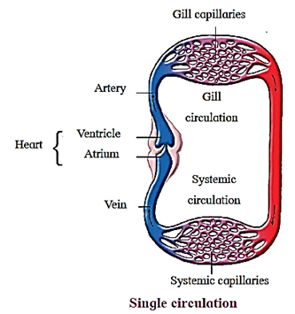
Closed circulation can be of two main types : single circulation and double circulation.

(a) Single circulation :

(a) Single circulation : In fishes heart shows single circulation as blood passes only once through heart during one cardiac cycle.

Deoxygenated blood is pumped from heart towards gills, where it undergoes oxygenation. This oxygenated blood moves towards various body parts, gets deoxygenated and returns back to heart for next cycle.

[collapse]

(b) Double circulation :

(b) Double circulation : Human heart shows double circulation as blood passes twice through the heart during one cardiac cycle.

  • The blood follows two routes, viz. pulmonary and systemic.
  • Pulmonary circulation is the circulation between the heart and the lungs. The course of blood during pulmonary circulation is from the right ventricle (by pulmonary trunk) to the left atrium (by two pairs of pulmonary veins) of heart through lungs.
  • Systemic circulation is the circulation between the heart and the body organs (except lungs). The course of blood during systemic circulation is from left ventricle (by systemic aorta) to all body organs and from the body back to right atrium (by vena cavae).
  • Coronary circulation is circulation to the cardiac muscles of the heart. Coronary arteries supply oxygenated blood whereas coronary veins join to form coronary sinus and collect deoxygenated blood. This sinus opens into the right atrium.

[collapse]

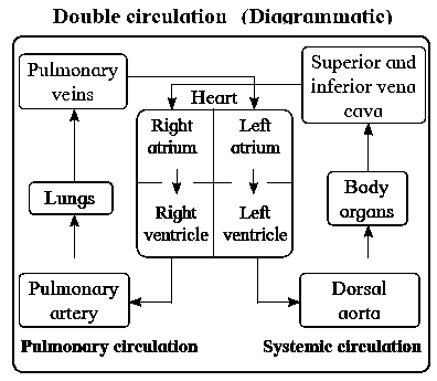
Circulatory System in Human :

  • Circulatory system in human is made up of blood vascular system and lymphatic system.
  • Blood vascular system consists of blood, heart and blood vessels.

 Blood composition and Coagulation :

  • Study of blood is called haematology.
  • The bright red, slightly alkaline main circulating, fluid in the human body is blood.
  • Blood is a fluid connective tissue derived from mesoderm. It has pH about 7.4.
  • There are about 5 litres of blood in the body which is about 8% of the total body weight.

Composition of blood : There are two main components of blood, viz., plasma (55%) and blood corpuscles (45%).

(i) Plasma : Plasma is a straw coloured fluid part of blood, slightly alkaline, viscous

fluid consisting of 90 — 92% water and 8 — 10% of solutes.

  • Solutes are 7% proteins (serum albumin, serum globulin, heparin, fibrinogen and prothrombin).
  • Other solutes are nutrients (glucose, amino acids, fatty acids and glycerol).
  • Nitrogenous wastes such as urea, uric acid, ammonia and creatinine.
  • Gases like oxygen, carbon dioxide and nitrogen.
  • Regulatory substances like enzymes and hormones.
  • Inorganic substances like bicarbonates, chlorides, phosphates and sulphates of sodium, potassium, calcium, magnesium, etc.

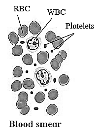
(ii) Blood corpuscles : Blood corpuscles are of three types, viz. erythrocytes (RBCs), leucocytes (WBCs) and thrombocytes (platelets).

Red blood corpuscles-erythrocytes :

Red blood corpuscles / erythrocytes :

  • Circular, biconcave, enucleated cells of about 7 /mm in diameter and 2.5 /mm in thickness.
  • RBC count is about 5.1 to 5.8 million RBCs/ cu mm in male and 4.3 to 5.2 million/cu mm in female. The average life span of RBC is about 120 days.
  • Erythropoiesis is formation of RBCs. It occurs in liver and spleen in foetus and in red bone marrow in adults.
  • The old RBCs are destroyed in liver and spleen.
  • Polycythemia is increase while erythrocytopenia is decrease in number of RBCs.
  • RBCs contain respiratory pigment called haemoglobin which helps in transport of oxygen and carbon dioxide.
  • The normal haemoglobin content in adult male is 14 — 17 gm/100 ml of blood and 13 - 15 gm/100 ml of blood in adult female.
  • Less amount of haemoglobin leads to anaemia.
  • RBCs transport oxygen from lungs to tissues and carbon dioxide from tissues to lungs. They maintain blood pH as haemoglobin acts as a buffer. They also maintain the viscosity of the blood.
  • RBCs also contains an enzyme, carbonic anhydrase.
  • The haematocrit is the ratio of the volume of RBCs to total blood volume of blood. Its Value is different in men and women.

[collapse]

White blood corpuscles/Leucocytes :

White blood corpuscles/Leucocytes :

  • Leucocytes are colourless, nucleated, amoeboid and phagocytic cells.
  • They show diapedesis, i.e. squeezing out of blood capillaries by amoeboid movement.
  • The size is about 8 to 15 um.
  • Total WBC count is 5000 to 11000 WBCs/ cu mm of blood.
  • The average life span of WBCs is about 3 to 4 days.
  • Leucopoiesis or formation of WBCs. It occurs in red bone marrow, spleen, lymph nodes, tonsils, thymus and Payer’s patches.
  • Leucocytosis is increase while leucopenia is decrease in the number of WBCs.
  • Leukaemia or blood cancer is a pathological increase in number of WBCs.
  • The dead WBCs are destroyed by phagocytosis in blood, liver and lymph nodes.
  • Leucocytes are of two types, viz., granulocytes and agranulocytes.
  • Granulocytes are of three types, viz. neutrophils, eosinophils and basophils.
  • Agranulocytes are of two types, viz. monocytes and lymphocytes.

[collapse]

Characteristics of Different Types of Leucocytes :

Characteristics of Different Types of Leucocytes :

Granulocytes : Granulocytes are cells with granular cytoplasm and lobed nucleus.

Based on their staining properties and shape of nucleus, they are of three types, viz. neutrophils, eosinophils and basophils.

(I) Neutrophils :

  • In neutrophils, the cytoplasmic granules take up neutral stains.
  • Their nucleus is three to five lobed.
  • It may undergo changes in structure hence they are called polymorphonuclear leucocytes or polymorphs.
  • Neutrophils are about 70% of total WBCs.
  • They are phagocytic in function and engulf microorganisms.

(II) Eosinophils or acidophils :

  • Cytoplasmic granules of eosinophils take up acidic dyes such as eosin. They have bilobed nucleus.
  • Eosinophils are about 3% of total WBCs.
  • They are non-phagocytic in nature.
  • Their number increases (i.e. eosinophilia) during allergic conditions.
  • They have antihistamine property

(III) Basophils :

  • The cytoplasmic granules of basophils take up basic stains such as methylene blue.
  • They have twisted nucleus.
  • In size, they are smallest and constitute about 0.5% of total WBCs.
  • They too are non-phagocytic.
  • Their function is to release heparin which acts as an anticoagulant and histamine that is involved in inflammatory and allergic reaction.

Agranulocytes : There are two types of agranulocytes, viz. monocytes and lymphocytes. Agranulocytes do not show cytoplasmic granules and their nucleus is not lobed. They are of two types, viz. lymphocytes and monocytes.

(I) Lymphocytes :

  • Agranulocytes with a large round nucleus are called lymphocyte.
  • They are about 30% of total WBCs.
  • Agranulocytes are responsible for immune response of the body by producing antibodies.

(II) Monocytes :

  • Largest of all WBCs having large kidney shaped nucleus are monocytes. They are about 5% of total WBCs.
  • They are phagocytic in function.
  • They can differentiate into macrophages for engulfing microorganisms and removing cell debris. Hence they are also called scavengers.
  • At the site of infections they are seen in more enlarged form.

[collapse]

Thrombocytes / Platelets :

  • Thrombocytes or platelets are non-nucleated, round and biconvex blood corpuscles.
  • They are smallest corpuscles measuring about 2.5 to 5 mm in diameter with a count of about 2.5 lakhs/cu mm of blood.
  • Their life span is about 5 to 10 days.
  • Thrombocytes are formed from megakaiyocytes of bone marrow. They break from these cells as fragments during the process of thrombopoiesis.
  • Thrombocytosis is the increase in platelet count while thrombocytopenia is decrease in platelet count.
  • Thrombocytes possess thromboplastin which helps in clotting of blood.
  • Therefore, at the site of injury platelets aggregate and form a platelet plug. Here they release thromboplastin due to which further, blood clotting reactions take place.

Blood clotting/ coagulation of blood : Active anticoagulants like heparin and antithrombin are present in the intact blood vessels. But upon the rupture of a blood vessel, bleeding starts. The fluid blood is converted into semisolid jelly by the process of blood coagulation or clotting.

The clotting of blood is a complicated process in which many factors (12 clotting factors) present in plasma and tissues are involved.

The event that take place during blood clotting :

The event that take place during blood clotting are as follows :

  • Release of thromboplastin from extrinsic source in tissue and intrinsic source in plasma at injured site through a step-Wise (cascade process) process.
  • Formation of enzyme prothrombinase in the blood.
  • Conversion of prothrombin into thrombin by prothrombinase.
  • Conversion of fibrinogen into fibrin by thrombin.
  • Formation of mesh by the fibrin fibres forming the clot.
  • The normal clotting time is 2 to 8 minutes.

[collapse]

Next Part->>

Click on link to get PDF from store

Rs 17

-Kitabcd Academy Offer-

Buy Notes(Rs.10)+ Solution(Rs.10) PDF of this chapter
Price : Rs.20 / Rs.17

Click on below button to buy PDF in offer (15% discount)

PDF :  Class 12th-Biology-Chapter-8-Respiration and Circulation-Text Book

PDF :  Class 12th-Biology-Chapter-8-Respiration and Circulation- Notes

PDF :  Class 12th-Biology-Chapter-8-Respiration and Circulation- Solutions

PDF SET :

All Chapters Notes-Class-12-Biology-(15-PDF)-Maharashtra Board-Rs-130

All Chapters Solutions-Class-12-Biology-(15-PDF)-Maharashtra Board-Rs-128

All Chapters Notes+Solutions-Class-12-Biology-(30-PDF)-Maharashtra Board-Rs-240

Useful Links

Main Page : – Maharashtra Board Class 12th-Biology All chapters notes, solutions, videos, test, pdf.

Previous Chapter : Chapter-7-Plant Growth and Mineral NutritionOnline Notes

Next Chapter : Chapter-9-Control and CoordinationOnline Notes

1 Comment

Add a Comment
  1. Nice notes it’s very helpful for me thank you kita abcd

Leave a Reply

Write your suggestions, questions in comment box

Your email address will not be published. Required fields are marked *

We reply to valid query.