Question 1.
Choose the correct option
(A) Location or position of meristematic regions is divided into ................... types
(a) one
(b) two
(c) three
(d) none of the above
(B) Cambium is also called ................
(a) apical meristem
(b) intercalary meristem
(c) lateral meristem
(d) none of the above
Answer :
(c) lateral meristem
[collapse]
(C) Collenchyma is a type of ................. tissue.
(a) living
(b) dead
(c) living and dead
(d) none of the above
(D) ..................... is a complex permanent tissue.
(a) Parenchyma
(b) Sclerenchyma
(c) Chlorenchyma
(d) Xylem
(E) Mesophyll tissue is present in................
(a) root
(b) stem
(c) leaf
(d) flower
Question 2.
Answer the following questions
(A) A fresh section was taken by a student but he was very disappointed because there were only few green and most colourless cells. Teacher provided a pink colour solution. The section was immersed in this solution and when observed it was much clearer. What is the magic?
Answer :
- The pink coloured solution given by teacher must be a safranin stain.
- Safranin is used to stain plant tissues, especially lignified tissues such as cell wall and xylem.
[collapse]
(B) While observing a section many scattered vascular bundles could be seen. Teacher said but in spite of this large number the stem cannot grow in girth. Why?
Answer :
- Students must have observed monocot stems.
- It is because, monocot stem shows scattered vascular bundles.
- In monocot stem, vascular bundles are closed 1.e. without cambium.
- Thus, secondary growth does not occur which is required for increase in girth.
Hence, in spite of having large number of scattered vascular bundles, monocot stems do not grow in girth.
[collapse]
(C) A section of the stem had vascular bundles, where one tissue was wrapped around the other. How will you technically describe it?
Answer :
- When one vascular tissue is completely encircling the other, it is called as concentric vascular bundle
- When phloem is encircled by xylem, it is called as leptocentric vascular bundle, whereas when xylem is encircled by phloem, it is called as hadrocentric vascular bundle.
- When xylem is encircled by phloem on both faces, it is called as amphicribral vascular bundle. When phloem is encircled by xylem on both faces it is called as amphivasal vascular bundle.
- Due to absence of cambium between xylem and phloem, concentric vascular bundles are always closed.
[collapse]
(D) There were two cut logs of wood lying in the campus. One had growth rings and other didn’t. Teacher said it is due to differences in their pattern of growth which is dependent on season. How?
Answer :
- It is possible that one of the cut logs was of a tropical tree, whereas the other was of a temperate tree. Since tropical trees grow in a similar manner all year, growth rings are not apparent. Another explanation for this could be that the log which had growth rings must be of an old tree which has experience many seasons, whereas the log without growth rings must be of younger tree, that has not been subjected to seasonal changes and hence not developed prominent growth rings.
- Growth rings are formed due to cambial activity during favourable and non-favourable climatic conditions.
- During favourable conditions, spring wood (early wood) is formed which has broader xylem bands, lighter colour, tracheids with thin wall and wide lumen, fibres are less in number, low density. Whereas, during unfavourable conditions, autumn wood (late wood) is formed which has narrow xylem band, darker in colour, lumen is narrow and walls are thick with abundant fibres, high density.
- Spring wood and autumn wood that appear as alternate light and dark concentric rings, constitute an annual ring or growth ring.
- These growth rings can be used to estimate the age of the tree. These are found more in older trees as compare to younger tree.
[collapse]
(E) While on the trip to Kashmir, Pintoo observed that cut portions of large trees shows distinct rings, which he never found in Maharashtra. Why is so?
Answer :
- Cut portions of large tress show distinct rings which are annual rings formed due to activity of cambium during favourable and non-favourable climatic conditions.
- Kashmir falls under temperate region where the climatic conditions are not unifomi through the year. In the spring season, conditions are favourable due to which cambium is active, whereas in autumn season, conditions are unfavourable due to which cambium is less active.
- This leads to formation of spring wood and autumn wood that appear as alternate light and dark concentric rings, constitute an annual ring or growth ring.
- Maharashtra falls under tropical region where climatic conditions are favourable throughout the year. In tropical areas, continuous growth of secondary xylem occurs. Thus, trees growing in tropical regions show less or no annual rings as compared to trees in temperate region.
[collapse]
(F) A student was observing a slide with no label under microscope. The section had some vascular bundles scattered in the ground tissue. It is section of a monocot stem! He exclaimed. No! it is section of fern rachis, said the teacher. Teacher told to observe vascular bundle again. Student agreed, Why?
Answer :
Let’s see the difference between fern rachis and monocot stem
| Fern rachis |
Monocot stem |
| In fern rachis, number of vascular bundles is less as compared to number of vascular bundles in monocot stem. |
In monocot stem, vascular bundles are numerous.
|
| In fern rachis, xylem consists of only tracheids |
In monocot stem, xylem consists of vessels (protoxylem and metaxylem) as well as tracheids. Monocot stem shows presence of lysigenous cavity just below protoxylem. |
| In fern rachis, phloem consists of only sieve cells |
In monocot stem, phloem consists of sieve tubes and companion cells. |
Thus, a student must have observed these differences in the given section and agreed to teacher’s statement that the given section is of fern rachis and not of monocot stem.
[collapse]
(G) Student found a wooden stopper in lab. He was told by an old lab attendant that it is there for many years. He kept thinking how it did not rot?
Answer :
- Wooden stopper or cork is obtained from the phellem (cork) part of a bark.
- Phellem (cork) is impervious in nature and does not allow entry of water due to suberized walls.
- Due to this it does not rot and remains as it is for many years.
[collapse]
(H) Student while observing a slide of leaf section observed many stomata on the upper surface. He thought he has placed slide upside down. Teacher confirmed it is rightly placed. Explain.
Answer :
- In a dicot leaf, stomata are generally absent on upper epidermis but are present on lower epidermis. Thus, the student must have thought that he has placed slide upside down.
- According to teacher, the section was placed rightly, thus the given section must be of monocot leaf.
- It is because, in monocot leaf stomata are present on both upper and lower epidermis.
[collapse]
Question 3.
Write short notes on the following points
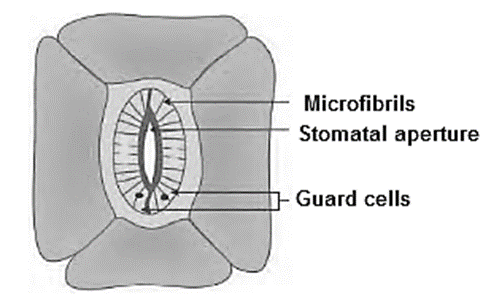
(A) Structure of stomata
Answer :
Structure of stomata :
- Small gateways in the epidermal cells are called as stomata.
- Stoma is controlled or guarded by specially modified cells called guard cells.
- These guard cells may be kidney shaped (dicot) or dumbbell shaped (monocot), collectively called as stomata.
- Guard cells have chloroplasts to carry out photosynthesis.
- Change in turgor pressure of guard cells causes opening and closing of stomata, which enables exchange of gases and water vapour.
- Stomata are further covered by subsidiary cells.
- Stoma, guard cells and subsidiary cells form a unit called stomatal apparatus.
[collapse]
(B) Secondary growth
Answer :
Secondary growth:
- Dicotyledonous plants and gymnosperms exhibit increase in girth of root and stem.
- In dicot stem, secondary growth begins with the formation of a continuous cambium ring.
- The cambium present between the primary xylem and primary phloem of a vascular bundle is called intrafascicular cambium.
- The cells of medullary rays adjoining these intrafascieular cambium strips become meristematic (regain the capacity to divide) and form the interfascicular cambium.
- Thus, a complete and continuous ring of vascular cambium is formed.
- The cambium ring cuts off new cells, towards both inner and outer sides.
- The cells that are cut-off towards pith (inner side) mature into secondary xylem and cells that are cut-off towards periphery mature into secondary phloem.
- Generally, amount of secondary xylem is more than the secondary phloem.
[collapse]
(C) Peculiarity of a sclerenchyma cell wall
Answer :
Peculiarity of a sclerenchyma cell wall:
- Cell wall of sclerenchyma is evenly thickened due to uniform deposition of lignin
- Cell wall of sclereids is extremely thick and strongly lignified.
[collapse]
Question 4.
Differentiate
(A) Vascular bundle of monocot and dicot
Answer :
| Vascular bundle of dicot |
Vascular bundle of monocot |
| Vascular bundle of dicot root :
1-Diarch, triarch or tetrarch condition can be observed. (Xylem bundles vary from two to six number)
2-Cambium is formed in later stage between xylem and phloem which causes secondary growtn
|
Vascular bundle of monocot root :
1-Polyarch condition is observed. (xylem bundles are more than six)
2-Secondary growth is absent.
|
| Vascular bundle of dicot stem :
1-Vascular bundles are arranged in the form of a ring.
2-It is conjoint, collateral and open (Cambium present)
3-Vascular bundle is not surrounded by a Sclerenchymatous bundle sheath.
4-Secondary growth takes place due to presence of cambium.
|
Vascular bundle of monocot stem :
1-Vascular bundles are scattered in the ground tissue.
2-They are conjoint, collateral and closed (cambium is absent).
3-Vascular bundle is surrounded by a
Sclerenchymatous bundle sheath.
4-Secondary growth does not occur due to absence of cambium.
|
| Vascular bundle of dicot leaf :
1-Vascular bundles of varying size.
2-The size of the vascular bundles is dependent on the size of the veins which vary in thickness in dicot leaf.
|
Vascular bundle of monocot leaf :
Vascular bundles are nearly of similar size (Except in main veins).
|
[collapse]
(B) Xylem and Phloem functioning
Answer :
| Xylem |
Phloem |
| Xylem conducts water and minerals from roots to the stem and leaves.
It also provides mechanical strength to the plant parts.
|
It is the chief food conducting tissue of vascular plants responsible for translocation of food from leaves to other plant parts. |
[collapse]
(C) Internal or anatomical difference between monocots and dicots.
Answer :
(1) Anatomy of dicots :
(i) Anatomy of dicot root :
- Pith is narrow.
- Diarch, triarch or tetrarch condition can be observed. (Xylem bundles vary from two to six umber)
- Cambium is formed in later stage between xylem and phloem which causes secondary growth.
(ii) Anatomy of Dicot stems :
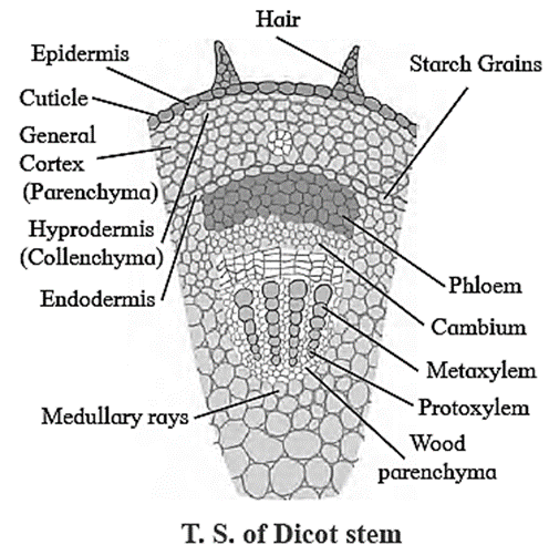
- Epidermis shows presence of multicellular trichomes.
- Hypodermis is made up of collenchymatous
- Medullary rays are present between vascular bundles.
- Vascular bundles are arranged in the form of a ring.
- It is conjoint, collateral and open (Cambium present).
- Vascular bundle is not surrounded by a sclerenchymatous bundle sheath.
- Secondary growth takes place due to presence of cambium.
- Pith is present.
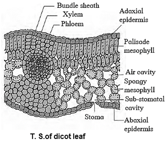
(iii) Anatomy of Dicot leaf :
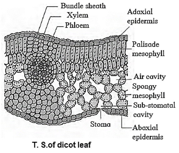
- Stomata : Stomata are restricted to lower epidermis. Guard cells of stoma are kidney shaped.
- Intercellular space : More intercellular spaces due to presence of spongy parenchyma,
- Venation : Reticulate venation.
- Vascular bundle : Vascular bundles of varying size. The size of the vascular bundles is dependent on the size of the veins, which vary in thickness in dicot leaf.
- Mesophyll cells : Mesophyll tissue is differentiated into palisade parenchyma and spongy parenchyma.
(2) Anatomy of Monocots :
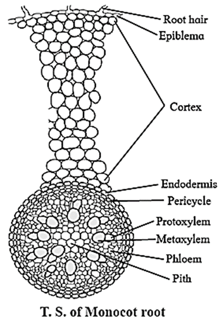
(i) Anatomy of monocot root :
- Pith is large and well developed.
- Polyarch condition is observed. (xylem bundles are more than six)
- Secondary growth is absent.
(ii) Anatomy of monocot stem :
- Epidermis is without trichomes.
- Hypodermis is made up of sclerenchymatous cells.
- Medullary rays are absent. :
- Vascular bundles are scattered in the ground
- They are conjoint, collateral and closed (cambium is absent).
- Vascular bundle is surrounded by a sclerenchymatous bundle sheath.
- Secondary growth does not occur due to absence of cambium.
- Pith is absent.
(iii) Anatomy of Monocot leaf :
- Stomata : Stomata occur on both epidermis. Guard cells of stoma are dumbbell shaped.
- Intercellular space : Less intercellular spaces as mesophyll is not differentiated into spongy and palisade tissue.
- Venation : Parallel venation.
- Vascular bundle : Vascular bundles are nearly of similar size (Except in main veins).
- Mesophyll cells : Mesophyll tissue is not differentiated into palisade parenchyma and spongy parenchyma.
[collapse]
Question 5.
Draw neat labelled diagrams
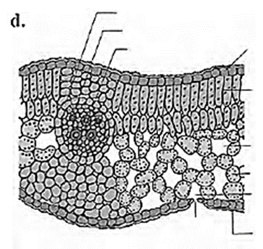
(A) T. S. of Dicot leaf.
Answer :
[collapse]
(B) T. S. of Monocot root.
Answer :
[collapse]
(C) T. S. of dicot stem.
Answer :
[collapse]
Question 6.
Write the information related to diagrams given below
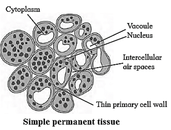
(A)
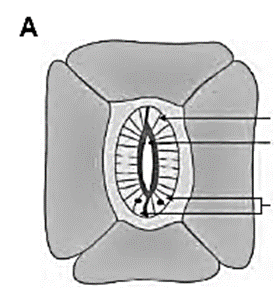
Answer :
The given diagram represents stoma in dicot leaf.
[collapse]
(B)
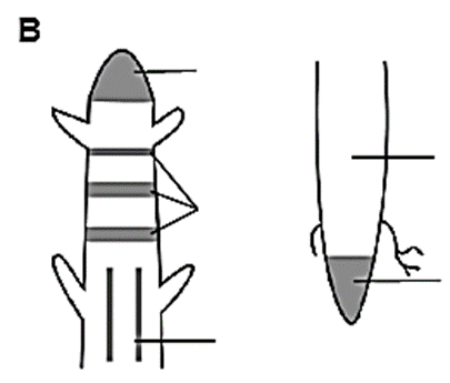
Answer :
Location of meristematic tissue
The labelled part can be considered as the ‘region of maturation’ of root apical however, the region of maturation does not contain meristematic tissue.
[collapse]
Question 7.
Identify the following diagrams, label it and prepare a chart of characteristics.
(a)

Answer :
The transverse section of a typical dicotyledonous root shows following anatomical features:
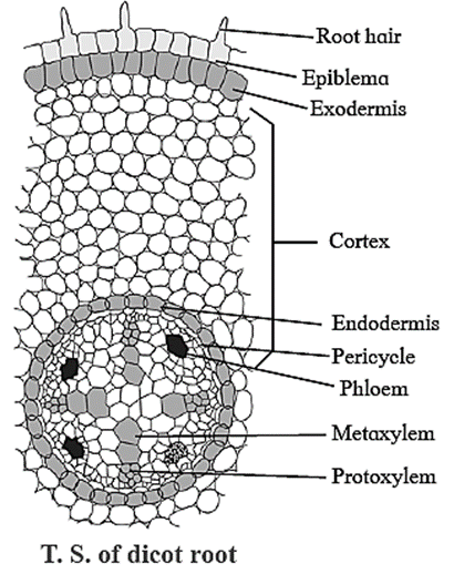
Epiblema: It is the outermost single layer of cells without cuticle. Some epidermal cells prolong to form unicellular root hairs.
Cortex: It is made up of many layers of thin walled parenchyma cells. Cortical cells store food and water.
Exodermis: After the death of epiblema, outer layer of cortex become cutinized and is called Exodermis.
Endodermis: The innermost layer of cortex is called Endodermis. The cells are barrel-shaped and their radial walls bear Casparian strip or Casparian bands composed of
suberin. Near the protoxylem, there are unthickened passage cells.
Stele: It consists of pericycle, vascular bundles and pith.
- Pericycle: Next to the endodermis, there is a single layer of thin walled parenchyma cells called pericycle. It forms outermost layer of stele or vascular cylinder.
- Vascular bundle: Vascular bundles are radial. Xylem and Phloem occur in separate patches arranged on alternate radii. Xylem is exarch in root that means protoxylem vessels are towards periphery and metaxylem elements are towards centre. Xylem bundles vary from two to six number, i.e. they may be diarch, triarch, tetrarch, etc.
- Connective tissue: A parenchymatous tissue is present in between xylem and phloem.
- Pith: The central part of stele is called pith. It is narrow and made up of parenchymatous cells, with or without intercellular spaces.
At a later stage cambium ring develops between the xylem and phloem causing secondary growth.
[collapse]
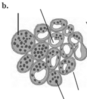
(b)
Answer :
The given figure represents simple permanent tissue i.e. Parenchyma.
Characteristics of parenchyma:
- It is a type of simple permanent tissue.
- Cells in this tissue are thin walled, isodiametric, round, oval to polygonal or elongated in shape.
- Cell wall is composed of cellulose.
- Cells are living with prominent nucleus and cytoplasm with large vacuole.
- Parenchyma has distinct intercellular spaces. Sometimes, cells may show compact arrangement.
- The cytoplasm of adjacent cells is interconnected through plasmodesmata and thus forms a continuous tissue.
- This is less specialized permanent tissue.
Occurrence:
- These cells are distributed in all the parts of a plant body viz. epidermis, cortex, pericycle, pith, mesophyll cells, endosperm, xylem and phloem.
[collapse]
Answer :
Features of stomata :
- Small gateways in the epidermal cells are called as stomata.
- Stoma is controlled or guarded by specially modified cells called guard cells.
- These guard cells may be kidney shaped (dicot) or dumbbell shaped (monocot), collectively called as stomata.
- Guard cells have chloroplasts to carry out photosynthesis.
- Change in turgor pressure of guard cells causes opening and closing of stomata, which enables exchange of gases and water vapour.
- Stomata are further covered by subsidiary cells.
- Stoma, guard cells and subsidiary cells form a unit called stomatal apparatus.
[collapse]
Answer :
Features of Dorsiventral Leaf :
- Dorsiventral Leaf is very common in dicotyledonous plants.
- In this mesophyll tissue is differentiated into palisade and spongy parenchyma.
- The leaves are commonly horizontal in orientation with distinct upper and lower surfaces. The upper surface which faces the sun is darker than the lower surface.
- Stomata is absent on the upper surface of these leaves.
[collapse]
Question 8.
Distinguish between Dicot and Monocot leaf on the basis of following characters.
| Characters |
Dicot leaf |
Monocot leaf |
| Stomata |
|
|
| Intercellular space |
|
|
| Venation |
|
|
| Vascular bundle |
|
|
| Mesophyll cells |
|
|
Answer :
| Characters |
Dicot leaf |
Monocot leaf |
| Stomata |
Stomata are restricted to lower epidermis. Guard cells of stoma are kidney shaped. |
Stomata occur on both epidermis. Guard cells of stoma are dumbbell shaped. |
| Intercellular space |
More intercellular spaces due to presence of spongy parenchyma, |
Less intercellular spaces as mesophyll is not differentiated into spongy and palisade tissue. |
| Venation |
Reticulate venation. |
Parallel venation. |
| Vascular bundle |
Vascular bundles of varying size. The size of the vascular bundles is dependent on the size of the veins, which vary in thickness in dicot leaf. |
Vascular bundles are nearly of similar size (Except in main veins).
|
| Mesophyll cells |
Mesophyll tissue is differentiated into palisade parenchyma and spongy parenchyma. |
Mesophyll tissue is not differentiated into palisade parenchyma and spongy parenchyma. |
[collapse]
We reply to valid query.