Notes-Part-1
|
Topics to be Learn : Part-1
|
Introduction :
Metabolism : An organism's body undergoes a variety of chemical reactions. The sum of these operations is known as metabolism.
Metabolic waste products : Metabolism produces a variety of by-products, some of which need to be eliminated. Such by-products are called metabolic waste products.
- Fluid, gaseous, organic, or inorganic waste products can result from metabolic processes.
- Metabolic wastes are produced inside body cells
Excretion and Excretory Products
Excretion : The process of eliminating waste products from the body is called excretion.
Various excretory products produced by the human body :
- Fluids such as water; gaseous wastes like CO2; nitrogenous wastes like ammonia, urea and uric acid, creatinine; minerals; salts of sodium, potassium, calcium, etc. if present in body in excess are excreted through urine, faeces and sweat.
- Pigments formed due to breakdown of haemoglobin like bilirubin (excreted through faeces) and urochrome (eliminated through urine).
- The pigments present in consumed foodstuffs like beet root or excess of vitamins, hormones and drugs.
- Volatile substances present in spices (eliminated through lungs).
- When does urine appear deeply coloured?
Ans : Urine can appear deeply coloured due to various reasons:
- Severe dehydration resulting in production of concentrated urine.
- Consumption of foodstuff like beet root, which contain coloured pigments
- Some medications can also cause the urine to appear deeply coloured.
Deamination :
- Deamination is the process of breakdown of excess amino acids.
- It is an essential process, since the body of an organism is unable to store excess amino acids.
- In this process, amino group is separated from the amino acid and ammonia is form.
- Toxic ammonia is either excreted or converted to less toxic forms like urea or uric acid before excretion.
Role of water in excretion :
- Ammonia is the basic product of deamination process.
- Ammonia is highly toxic and needs to be diluted immediately.
- If there is no or limited access to water, need for conversion of ammonia becomes necessary.
- Hence, the availability of water plays a key role in deciding the mode of excretion of an organism.
Three main modes of excretion in animals : The three main modes of excretion in animals are as follows: (i) Ammonotelism (ii) Ureotelism (iii) Uricotelism (i) Ammonotelism: (ii) Ureotelism : (iii) Uricotelism:
Sharks retain more urea in their blood :
Reason : Sharks retain more urea in their body fluid (blood) to make their blood isotonic to surrounding marine water (in order to maintain osmotic balance). This helps them to prevent possible loss of water by exosmosis.
Terrestrial animals are generally either ureotelic or uricotelic, not ammonotelic :
Reason : Ammonia is highly toxic to animals. An animal requires large amount of water to dissolve and eliminate ammonia. Terrestrial animals cannot lose such a large amount of water. Ureotelic and uricotelic animals require less amount of water for removal of nitrogenous waste, to conserve water, ureotelism and uricotelism is adapted by terrestrial animals.
Guanotelic Animals : Animals like spiders, scorpions and penguins are guanotelic organisms as they excrete guanine.
Plasma creatinine : Plasma creatinine is a waste product produced by muscles from the breakdown of a compound called creatine phosphate’
- Plasma creatinine is produced from catabolism of creatinine phosphate during skeletal muscle contraction.
- It provides a ready source of high energy phosphate.
- Normally blood creatinine levels remain steady because the rate of production matches its excretion in urine.
- Hence, plasma creatinine is used as an index of kidney function and its level above normal is an indication of poor renal function.
Homeostasis :
- Homeostasis is the maintenance of constant internal environment of the body.
- Homeostasis is however dependent on osmoregulation, which is the process of controlling solute concentrations and water balance.
- The composition of blood and therefore the internal environment is highly dependent on what the excretory organs retain in the body.
- Hence, excretion plays an important role in homeostasis.
Osmoregulation :
Osmoregulation is the process of maintaining an internal concentration of salt and water in the body of fishes.
Osmoregulation in Freshwater fishes:
- The salt concentration inside the body of freshwater fishes is higher than their surrounding water. Due to this, water enters the body due to osmosis.
- If the flow of water into the body is not regulated, fishes would swell and get bigger.
- To compensate this, the kidneys produce a large amount of urine.
- Excretion of large amounts of urine regulates the level of water in the body but results in the loss of salts.
- Thus, in order to maintain a sufficient salt level, special cells in the gills (chloride cells) take up ions from the water, which are then directly transported into the blood.
Osmoregulation in Marine fishes:
- Since the salt content in blood of marine fishes is much lower than that of seawater, they constantly tend to lose water and build up salt.
- To replace the water loss, they continually need to drink seawater.
- Since their small kidney can only excrete a relatively small amount of urine, salt is additionally excreted through gills, where chloride cells work in reverse as in freshwater fishes.
Osmoregulation in Marine organisms :
- Marine birds like Albatross have special glands called salt glands near their nostrils.
- These glands are capable of secreting salts by active transport and help to manage osmotic balance
- The salt glands in Albatross are located in or on the skull in the area of eyes.
- Examples of organisms that possess salt glands are birds like Albatross, marine organisms like sea turtles, marine iguanas, sea birds, gulls, etc.
Osmoconformers and Osmoregulators :
- Animals can either be isoosmotic to the surrounding (osmoconformers) or control internal environment independent of external environment (osmoregulators).
- Marine organisms mostly are osmocomformers because their body fluids and external environment are isoosmotic in nature.
- Fresh water forms and terrestrial organisms are osmoregulators.
Methods of excretion in various organisms :
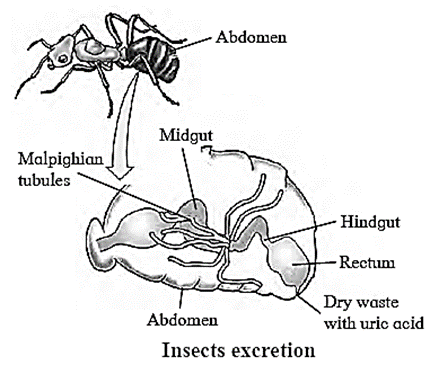
Nephridia : Nephridia is a simple or branching tubule used for excretion which opens to the exterior through pores called nephridiopores.
Two major types of nephridia are as follows:
- Protonephridia: These are network of dead end tubes called flame cells. They are mostly found in animals that lack a true body cavity. e. g. Platyhelminthes, rotifers, some amelids and Amphioxus.
- Metanephridia: These are unbranched coiled tubes that are connected to the body cavity through funnel like stmctures called nephrostomes. Body fluid enters the nephridium through nephrostome and gets discharged through nephridiopore. e.g. Earthworms.
Excretory System in Human Being
Excretory system of man consists of; (i) Kidneys (ii) Ureters (iii) Urinary Bladder (iv) Urethra
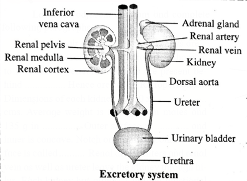
(i) Kidneys :
Structure of Kidney:
- A pair of bean shaped kidneys are present on either side of the backbone from 12th thoracic to 3rd Lumbar vertebra.
- Kidneys are present behind peritoneum. Hence are called Retroperitoneal. Dimensions of each kidney are 10 x 5 x 4 cms. Average weight is 150 g in males and 135 g in females.
- Outer surface is convex and inner is concave. Notch on the inner concave surface is called hilum.
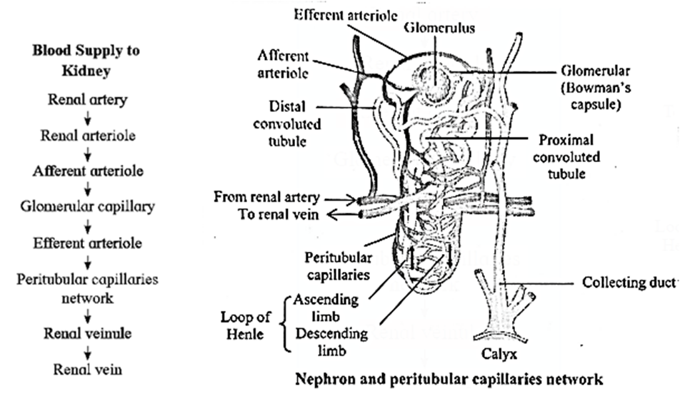
- Renal artery enters and renal vein as well as ureter leave the kidney through hilus. Each kidney has almost 1 million functional units called nephron.
Function of Kidney:
- Kidneys bring about separation and elimination of nitrogenous waste, excess water and toxic substances from the body.
- They play a role in osmoregulation and regulation of pH of body fluids and thus maintain body homeostasis.
- They produce calcitriol and renin.
- They secrete eiythropoietin which is essential for production of RBCs.
(ii) Ureters:
Structure of Ureters:
- A pair of ureters arise from hilum of each kidney. Each ureter is a long muscular tube 25 — 30 cm in length.
- Ureters open into urinary bladder by separate openings, which are not guarded by valves.
- They pass obliquely through the wall of urinary bladder. This helps in prevention of backward flow of urine due to compression of ureters while bladder is filled.
Function of Ureters:
- Ureters transport urine from renal pelvis to urinary bladder.
(iii) Urinary bladder:
Structure of Urinary bladder:
- It is a median pear-shaped sac. A hollow muscular organ, the bladder is situated in pelvic cavity posterior to pubic symphysis.
- At the base of the urinary bladder there is a small inverted triangular area called Trigone.
- At the apex of this triangle is opening of urethra. At the two points of the base of the triangle are openings of ureters.
- Urinary bladder is covered externally by peritoneum.
- Inner to peritoneum is muscular layer. It is formed by detrusor muscles which consist of three layers of smooth muscles. Longitudinal - circular - longitudinal respectively.
- Innermost layer is made up of transitional epithelial tissue. It helps bladder to stretch.
Function of Urinary bladder :
- Urinary bladder is a temporary storage organ for urine. It helps to expel urine (micturition).
(iv) Urethra:
Structure of Urethra:
- It is a fibromuscular tube-like structure arising from urinary bladder and opening to the exterior of the body.
- There are two urethral sphincters between urinary bladder and urethra.
- Internal sphincter: Made up of detrusor muscles, involuntary in nature.
- External sphincter: Made up of striated muscles, voluntary in nature.
- If this valve is not functioning properly during inflammation of bladder, it can lead to kidney infection.
- The urethra in women (4 cm) is much shorter than that of males (20 cm). This allows easy passage of bacteria into the urinary bladder. Hence, urinary tract infections are more common in females than males.
Function of Urethra:
- Urethra is a passage way for discharging urine from body. In males, it acts as urinogenital organ.
| Know This :
Floating Kidney :
|
Micturition :
- Micturition is the process of releasing urine from the urinary bladder.
- The typical urine bladder capacity is 700 ml.
- When the urinary bladder is nearly half full, stretch receptors in the bladder send signals to the spinal cord, causing a conscious desire to release pee.
- The spinal cord's micturition reflex centre sends signals to the urinary bladder wall and the internal urethral sphincter.
- Bladder muscles tighten and internal urethral sphincter muscles relax.
- The external sphincter relaxes after receiving impulses from the conscious centre of the brain.
- This results in urine elimination from the bladder.
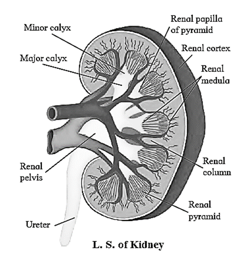
L.S of kidney : Each kidney has three layers of tissue covering it: the outermost renal fascia, the middle adipose capsule, and the innermost renal capsule. The L.S, of kidney shows two distinct regions within the capsule. Histologically, kidney is divisible into two regions as renal cortex and renal medulla.
Nephrology : It is a branch of biology that deals with the structure, function and disorders of male and female urinary system.
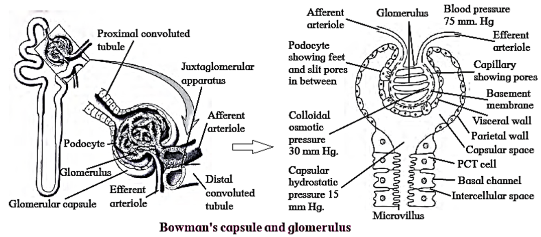
Nephrons :
- Nephrons are structural and functional units of kidney.
- Each nephron consists of a 4-6 cm long, thin-walled tube called the renal tubule and a bunch of capillaries known as the glomerulus.
- The wall of the renal tubule is made up of a single layer of epithelial cells.
- Its proximal end is wide, blind, cup-like and is called as Bowman’s capsule, whereas the distal end is open.
- The nephron is divisible into Bowman’s capsule, neck proximal convoluted tubule (PCT), Loop of Henle (LoH), distal convoluted tubule (DCT) and collecting tubule (CT).
- The glomerulus is present in the cup-like cavity of B0wman’s capsule and both are collectively known as renal corpuscle or Malpighian bodv.
Structure of nephron : Each nephron is divided into two main parts: (i) Malpighian body (ii) Renal tubule (i) Malpighian body: Each Malpighian body is about 200um in diameter and consists of a Bowman’s capsule and glomerulus. Glomerulus: Bowman’s capsule: [Podocytes are a special type of squamous cells that have a foot-like pedicel. They are present in the visceral wall of the Bowman’s capsule and are in close contact with the walls of capillaries of glomerulus] (ii) Renal tubule: Neck: Proximal Convoluted Tubule : Loop of Henle : Distal convoluted tubule: Collecting tubule:
Cortical nephrons and Juxtamedullary nephrons :
Cortical nephrons :
- They have a shorter loop of Henle.
- Loop of Henle of these nephrons extends very little into the medulla. .
- Most nephrons are crotical nephrons.
- Efferent arteriole forms peritubillar capillary network around DCT, PCT and Henle’s loop of cortical nephrons
Juxtamedullary nephrons :
- They have a longer loop of Henle.
- Loop of Henle of these nephrons run deep into the medulla.
- Few nephrons are Juxtamedullary nephrons.
- Efferent arteriole forms loop-shaped vasa recta around Henle’s loop of juxtamedullary nephrons.
Juxta Glomerular Apparatus :
- Some smooth muscle cells of the wall of afferent arteriole are modified in such a way that their sarcoplasm is granular. These cells are called juxtaglomerular (JG) cells.
- In each nephron, initial part of DCT makes contact with the afferent arteriole of same nephron.
- Cells in the wall of DCT in this region are packed more densely than those in other region of DCT. This is called macula densa.
- Macula densa and the JG cells together form Juxta Glomerular Apparatus (JGA).
- The JGA plays an important role in blood pressure regulation within the kidney.
We reply to valid query.