|
Topics to be Learn : Part-1
- Introduction
- Nervous Coordination
- Nervous System in Hydra
- Nervous System in Planaria (flatworm)
- Neural tissue
- Synapse
- Transmission of nerve impulse along the axon
- Human Nervous System
- Sensory Receptors
- Disorders of nervous system
Topics to be Learn : Part-2
- Chemical Coordination
- Endocrine system
- Major endocrine glands
|
-Chemical Coordination-
The cells in organisms communicate with each other through chemical signals. These cells are broadly of four types as follows :
- Autocrines : Cells release secretion to stimulate themselves.
- Paracrines : Cells release secretion to stimulate neighbouring cells.
- Endocrines : Cells release secretion to stimulate distant cells.
- Pheromones : Cells/Organs release secretions to stimulate other organism.
Chemical coordination is carried out by secretions of ductless glands or endocrine glands. Hence this chemical coordination system is also called the endocrine system.
Endocrine system :
- The endocrine system controls body activities by means of chemical messengers called hormones.
- Endocrine glands are ductless glands which are capable of secreting hormones.
- The hormones are poured directly into the bloodstream as the endocrine glands do not have duct.
- Hormones regulate the function of target tissue or organ.
- They either have excitatory effect or have an inhibitory effect.
Main endocrine glands in human body :
Main endocrine glands in human body :
The main endocrine glands in human body are as follows :
- Pituitary or hypophysis
- Hypothalamus
- Thyroid
- Parathyroid
- Adrenal or suprarenal
- Islets of Langerhans in pancreas
- Endocrine parts of gonads, i.e. testis and ovary.
Pineal gland and thymus are also endocrine glands of less importance.
[collapse]
Properties of Hormones :
- They act as chemical messengers and are effective in very low concentration.
- Hormones can function as regulators that inhibit or stimulate or modify specific processes.
- Hypersecretion or Hyposecretion of hormones leads to various disorders.
- These are metabolised after their function and are excreted through urine.
Mechanism of hormone action :
Mechanism of hormone action :
- Hormones are released in a very small quantity.
- They produce their effect on the target organs/cells by binding to hormone receptors.
- The hormone receptors may be on the cell membrane or may be intracellular.
- A hormone receptor complex is formed and this leads to biochemical change in the target tissue.
Mode of hormone action through membrane receptors :
- Hormones like catecholamines, peptide and polypeptide hormones are not lipid soluble.
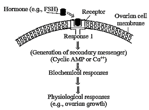
- Therefore they cannot enter their target cells through plasma membrane.
- Molecules of amino acid derivatives, peptide hormones bind to specific receptor molecules located on the plasma membrane.
- The hormone receptor complex causes the release of an enzyme adenylate cyclase from the receptor site. This enzyme forms cyclic AMP from ATP of the cell.
- The hormone acts as the first messenger and cAMP is the second messenger.
Mode of hormone action through intracellular receptors :
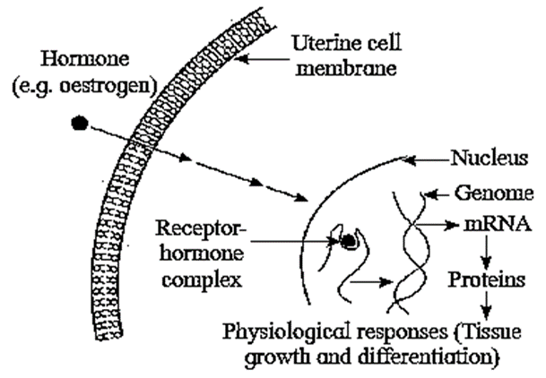
- Steroid and thyroid hormones are lipid soluble and easily pass through plasma membrane of target cell into the cytoplasm.
- In the cytoplasm, they bind to specific intra-cellular receptor proteins forming a hormone-receptor complex that enters the nucleus.
- In the nucleus, the hormone receptor complex binds to a specific regulatory site of DNA.
[collapse]
Q. Hormones are called chemical messenger, and regulators. Explain.
Answer :
- Hormones bring about coordination in the body with the help of nervous system.
- Endocrine system and nervous system together form neuro-endocrine system.
- This system works in tune with the external and internal environmental changes.
- The hormones are either excitatory or inhibitory. They bring about the actions accordingly to keep the body in homeostasis or equilibrium.
- Almost all endocrine glands are controlled by negative feedback inhibition. Some glands are auto-regulatory. Therefore, the concentration of hormones cannot be in excess or in deficiency.
- Almost all the functions such as metabolism growth, reproduction, etc. are under the control of hormones. Therefore hormones are called regulators and messengers.
[collapse]
Major endocrine glands
Hypothalamus :
- Ectodermal in origin.
- Forms the floor of diencephalon.
- Major function is to maintain homeostasis.
- Controls the secretory activity of pituitary gland by the release and inhibiting hormones.
- All hormones of hypothalamus are peptide hormones.
Hormones of hypothalamus :
Hormones of hypothalamus :
- Somatotropin/GHRF
- Somatostatin/GHRIF
- Adrenocorticotropin Releasing Hormone
- Thyrotropin Releasing Factor
- Gonadotropin Releasing Hormone (GnRH)
- Prolactin Inhibiting Hormone (Prolactostatin)
- Gastrin Releasing Peptide (GRP)
- Gastric Inhibitory Polypeptide (GIP)\
[collapse]
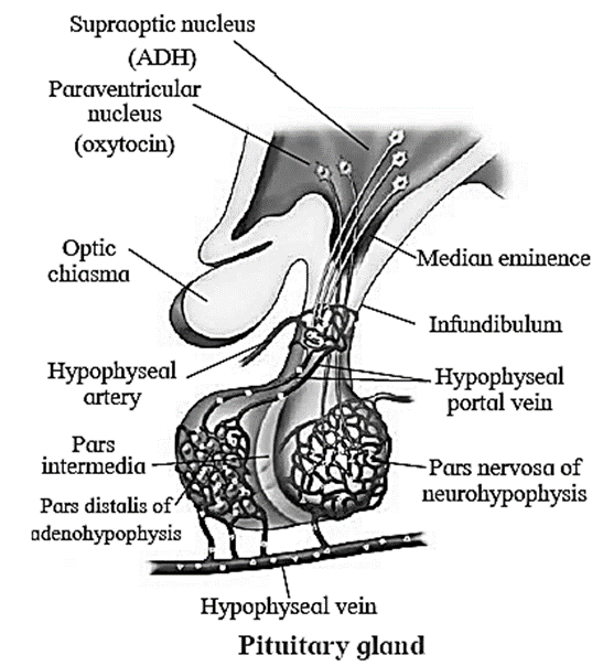
Pituitary gland or hypophysis :
External morphology :
- Pea sized reddish-grey coloured gland.
- Controls almost all other endocrine glands, hence previously it was called the master endocrine gland.
- However, hypothalamus controls it through the releasing and inhibiting factors.
- Located just below the hypothalamus and is attached to it by a stalk called infundibulum or hypophyseal stalk.
- Remains lodged in a bony depression called sella turcica of the sphenoid bone.
- Consists of two lobes called anterior lobe (Adenohypophysisl and posterior lobe
- (Neutohypophysis). Intermediate lobe (Pars intermedia) is a small reduced part lying in the cleft between the anterior and posterior lobe.
- Neurohypophysis is connected directly to the hypothalamus by axon fibres forming hypothalamo-hypophyseal tract,
- Adenohypophysis and intermediate lobe are connected to the hypothalamus through hypothalamo-hypophyseal portal system.
Parts, morphology, histology and functions of pituitary :
Parts, morphology, histology and functions of pituitary :
The pituitary gland shows two distinct regions : Anterior lobe or adenohypophysis and posteriorlobe or neuro-hypophysis.
(1) Adenohypophysis or Anterior lobe : It is the largest lobe of the gland and forms about 75% of pituitary gland. It develops as an outgrowth called Rathke‘s pouch from the roof of embryonic buccal cavity. It has three divisions, viz. pars tuberalis, pars distalis and pars intermedia.
- (i) Pars tuberalis :Tubular region present below the hypothalamus is known as pars tuberalis. It is like a collar around the infundibulum. It is non-secretory in nature.
- (ii)Pars distalis : The largest anterior region which is secretory in nature is called pars distalis. It is made up of loose cords of epitheloid secretory cells which are separated by reticular connective tissue containing blood sinusoids. It is connected to the hypothalamus by portal system formed by blood sinusoids.
- Histology of Pars distalis : Secretory epithelial cells with blood capillaries. Has chromophobe and chromophil cells. Chromophil cells are of two types, viz. acidophils and basophils. Acidophils are of two types, viz. somatotropes and lactotropes. Basophils are of three types, viz. thyrotropes, gonadotropes and corticotropes.
- Functions of Pars distalis : Secrete GH, TSH, ACTH, LTH, GTH (FSH, LH/ICSH)
- (iii) Pars intermedia : The narrow cleft between the pars distalis and pars nervosa of neuro-hypophysis is called the intermediate part or pars intermedia.
- Histology of Pars intermedia : Not well defined in man
- Functions of Pars intermedia : It is reduced, less developed and non-functional in human being. Larger and functional in lower vertebrates
(2) Neuro-hypophysis or Posterior lobe : The posterior lobe of the pituitary which is attached to hypothalamus by infundibular stalk is called neuro-hypophysis. It is smaller and constitute 25% of pituitary. It has the following three parts :
- Median eminence : The swollen median part of the hypothalamus where infundibulum gets attached is called median eminence.
- Infundibulum : Infundibulum is the hypophyseal stalk that helps in attachments of pituitary gland to the hypothalamus contains mainly the axonic fibres of neurosecretory cells present in hypothalamus. Functions : It forms the major connection of hypothalamo-hypophysis axis.
- Pars nervosa : The lowermost, larger region of neuro-hypophysis that contains axons is called pars nervosa. It acts as a neurohaemal organ and contains specialized cells caged pituicytes.
- Histology of Pars nervosa: Pituicytes and axonic knobs (Herring's bodies)
- Functions of Pars nervosa : Store ADH and Oxytocin. It is neurohaemal organ.
[collapse]
Hypothalamo - Hypophyseal portal system :
- Various hormones secreted by hypothalamus reach the pituitary gland through this portal system.
- The portal vein collects blood from various parts of hypothalamus and opens into anterior lobe of pituitary.
- From pituitary, the vein finally carries the blood into the superior vena cava.
Hormones of pituitary and their role :
Hormones of pituitary and their role :
(i) Somatotropin or Somatotropic hormone (STH) or growth hormone (GH) :
Cells secreting the hormone - Acidophils of adenohypophysis (Somatotropes).
Regulation :
- Dual control of hypothalamus GHRF and GHIF or somatostatin
Function :
- General growth of the body
Disorders :
- Hyposecretion : Dwarfism; pituitary infantilism in childhood (Frohlic dwarfs-mentally abnormal and Lorain dwarfs-mentally normal). Also called midgets. Simrnond’s disease in adulthood.
- Hypersecretion : Gigantism in childhood, Acromegaly in adulthood.
(ii) Thyrotropin or Thyroid stimulating hormone (TSH) :
Cells secreting the hormone -Basophils of adenohypophysis (Thyrotropes)
Regulation :
- TRF from hypothalamus. Negative feedback between level of thyroxine and TSH.
Function :
- Stimulates thyroid gland and helps in the formation of thyroxine.
Disorders :
- Hyposecretion : Thyroid atrophy.
- Hypersecretion : Increased BMR, loss of weight and increased heart rate and blood pressure.
(iii) Corticotropin or Adrenocorticotropic hormone (ACTH) :
- Cells secreting the hormone : Basophils called corticotrophs
Regulation :
- CRF from hypothalamus. Negative feedback between level of cortisol and ACTH.
Function :
- Stimulates adrenal cortex and helps it to secrete glucocorticoids and mineralocorticoids.
Disorders :
- Hyposecretion : Rheumatoid arthritis, Addison’s disease.
- Hypersecretion : Cushing’s disease.
(iv) Follicle stimulating hormone (FSH) :
- Cells secreting the hormone : Basophils
Regulation :
- Regulation by gonadotropin releasing factors of hypothalamus. Negative feedback between the sex hormones- especially estrogen and FSH.
Function :
- In females : Stimulation of germinal epithelium of ovary, oogenesis, stimulation of ovarian follicular cells to secrete estrogen. Estrogen helps in the development of secondary sexual characters in female.
- In males : Stimulation of seminiferous tubules and spermatogenesis maturation of sperms. Hyposecretion or deficiency causes infertility in both the sexes.
(v) Luetinizing hormone (LH in females) :
- Cells secreting the hormone : Basophils
Regulation :
- Negative feedback between LH and progesterone.
Function :
- Ovulation formation of corpus leuteum, stimulation of corpus leuteum to produce progesterone. Maintenance of pregnancy.
(vi) Interstitial Cell Stimulating Hormone (ICSH in males) :
- Cells secreting the hormone : Basophils
Regulation :
- Negative feedback between ICSH and testosterone.
Function :
- Stimulates interstltlal cells of Leydig stimulation to secrete testosterone, Testosterone develops secondary sexual characters in males.
(vii) Luteotropic hormone or Prolactin LTH Or PL :
- Cells secreting the hormone : Acidophils of adenohypophysis
Regulation :
- PRF and PIF from hypothalamus regulate it.
Function :
- Mammotropic : Development of mammary glands.
- Lactogenic : Milk secretion.
- Luteotropic : Maintenance of corpus luteum, Stimulation of corpus luteum to produce progesterone.
(viii) Melanocyte Stimulating Hormone (MSH) :
- Cells secreting the hormone : Pars intermedia
Regulation :
- Hypothalamic melanostatin
Function :
- Stimulation of melanocytes of skin and synthesis of melanin, darkening the skin.
(viii) Antidiuretic hormone (ADH or Vasopressin) :
Cells secreting the hormone :
- Hypothalamic neurons secrete and neuro-hypophysis stores these two hormones.
- Neuro-hypophysis is called neuro-haemal organ due to such storage.
Regulation :
- Regulation through osmotic pressure of the blood.
- Osmoreceptors in hypothalamus thus indirectly regulate ADH secretion.
Function :
- Increasing permeability of distal convoluted tubule in nephron.
- Facilitates ultrafiltration in kidneys. Decreases urine output.
- Constricts arterioles and increases blood pressure.
Disorders :
- Hyposecretion : Diabetes insipidus—causes polydipsia and polyuria.
- Hypersecretion : Antidiuresis and water retention in the body.
(ix) Birth hormone (Oxytocin) :
Regulation :
- Secreted in large amount during childbirth or parturition
Function :
- Contraction of uterine myometrium during parturition.
- Ejection of milk through mammary glands.
- Ascent of spermatozoa during copulation.
- Excitation of musculature of gall bladder, ureters, urinary bladder and intestine.
(x) Coherin : Function -Induce contractions of jejunum
[collapse]
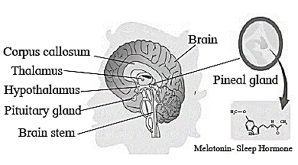
Pineal gland :
- The pineal body/pineal gland is given off from the roof of diencephalon. It is located between the two cerebral hemispheres.
- The pineal gland is sensitive to the biochemical signals of light.
- It secretes a hormone called melatonin also known as sleep hormone.
Thyroid gland :
Morphology :
- It is the largest endocrine gland.
- The two lobes of thyroid gland are connected a non-secretory band called isthmus.
Internal structure :
Internal structure :
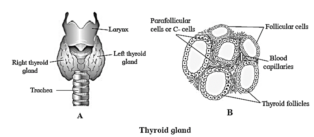
- The thyroid lobes are composed of rounded follicles held together by inter follicular connective tissue called stroma.
- The stroma contains blood capillaries and small group of parafollicular cell or ‘C’ cells.
- Thyroid follicles are composed of cuboidal epithelium resting on a basement membrane and is filled with a gelatinous colloid.
[collapse]
Thyroid hormones :
- The two hormones secreted by the follicular cells are Thyroxine/tetra iodothyronine/T4 (four atoms of iodine) and Triiodothyronine or T3 (three atoms of iodine).
- Parafollicular cells produce a hormone thyrocalcitonin whose production is not under the control of TSH.
Formation of T3 and T4 :
- Thyroxine is synthesized by attaching iodine to amino acid tyrosine by enzymatic action.
- The amino acid tyrosine molecule binds to iodine to produce Monoiodotyronine (T1) or 2 atoms of iodine to produce Diiodothyronine (T2).
- T1 and T2 molecules bind end to end to make colloidal mass inside the follicle. They are further metabolised to prepare T3 and T4.
Functions of Thyroid hormones :
Functions of Thyroid hormones :
- Regulation of the basal metabolic rate of body
- Regulation of metabolism by stimulating protein synthesis and promotes growth of body tissues.
- Calorigenic effect as it helps in thermoregulation by increasing heat production.
- Increases action of neuro transmitters —adrenaline and nor adrenaline.
- Supports the process of RBC production and maintenance of Water and electrolyte balance.
- Regulates reproductive cycles in females.
- Parafollicular cells or ‘C’ cells produce thyrocalcitonin hormone, which regulates calcium metabolism.
- Calcitonin is the active form of hormone, which is hypocalcemic hormone. It regulates the concentration of calcium and phosphorus in the blood.
[collapse]
Functional disorders of thyroid gland :
Functional disorders of thyroid gland :
Disorders of thyroid gland are of three types, viz. hypothyroidism, hyperthyroidism and simple goitre.
Hypothyroidism : Hypothyroidism is deficient secretion of thyroxine. This hyposecretion causes two types of disorders, viz. cretinism in children and myoxedema adults.
- Cretinism : Hyposecretion of thyroxine ml childhood causes cretinism. The syrnptoms of cretinism are retardation of physical and mental growth.
- Myxoedema : Deficiency of thyroxine in adults causes this disorder. It is also referred to as Gull’s disease. Symptoms are thickening and puffiness of the skin and subcutaneous tissue particularly of face and extremities. Patients with low BMR. It also causes mental dullness, loss of memory, slow action.
Hyperthyroidism : Excessive secretion of thyroxine causes exophthalmic goitre or
Grave’s disease. There is slight enlargement of thyroid gland. It increases BMR, heart rate, pulse rate and BR Reduction in body weight due to rapid oxidation, nervousness, irritability. Peculiar symptom is exophthalmos, i.e. bulging of eyeballs with staring look and less blinking. This is caused by deposition of fats behind the eye balls in eye sockets. There is muscular weakness and loss of weight.
Simple goitre (Iodine deficiency goitre) : Simple goitre occurs due to deficiency of iodine in diet or drinking water. Simple goitre causes enlargement of thyroid gland. Thyroid gland in an attempt to get more iodine from the blood, swells due to increased blood supply. Prevention of goiter can be done by administering iodized table salt. It is also called endemic goitre as it is common in hilly areas.
[collapse]
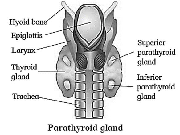
Parathyroid gland :
Parathyroid gland :
- Situated on the posterior surface of the lobes of thyroid gland.
- 2 pairs, named as superior and inferior parathyroid glands.
- Cells are arranged in a compact mass.
Hormones :
- The parathyroid secretes a peptide hormone called parathormone [PTH). It is also called Collip’s hormone.
- Regulates calcium and phosphate balance between blood and other tissues. It is a hypercalcemic hormone. Release of parathormone increases blood calcium level.
- It stimulates osteoclast of bones to stimulate bone resorption.
- Thus, parathormone and calcitonin are antagonistic hormones.
Disorders :
- Hyposecretion of parathormone lowers concentration of calcium in the blood. This increases excitability of nerves and muscles causing muscle twitch and spasm. This is called parathyroid tetany or hypocalcaemic tetany.
- Hypersecretion of parathormone is responsible for more absorption of calcium from bones i.e., demineralization of bones resulting insoftening, bending and fracture of bone. This is called osteoporosis. It is common in women who have reached menopause.
[collapse]
Thymus gland :
Thymus gland :
- Located in the upper part of thorax on the dorsal side of the heart just behind sternum.
- Prominent gland at birth till puberty but gets gradually atrophied in the adult due to action of sex hormones.
Functions:
- Secretes the hormone thymosin.
- Important role in the development of immune system by maturation of T-lymphocytes
[collapse]
Adrenal gland/Suprarenal gland :
- Adrenal glands have dual origin from mesoderm and ectoderm.
- They are located on the upper border of each kidney.
- Adrenal glands are small, conical yellowish glands and show two distinct regions, outer cortex and inner medulla.
| (i) Adrenal cortex (outer) |
(ii) Inner medulla |
| -Adrenal cortex is derived from embryonic mesoderm.
-Adrenal cortex secretes many hormones together called corticoids.
-It is differentiated into three concentric regions.
|
-Derived from embryonic ectoderm.
Secretes main two hormones
(1) Adrenaline (epinephrine)- (Emergency hormone, also called 3F hormone — (fight, flight and fright).
(2) Noradrenaline (norepinephrine)- [Regulates the blood pressure under normal condition, acts as vasoconstrictor)
|
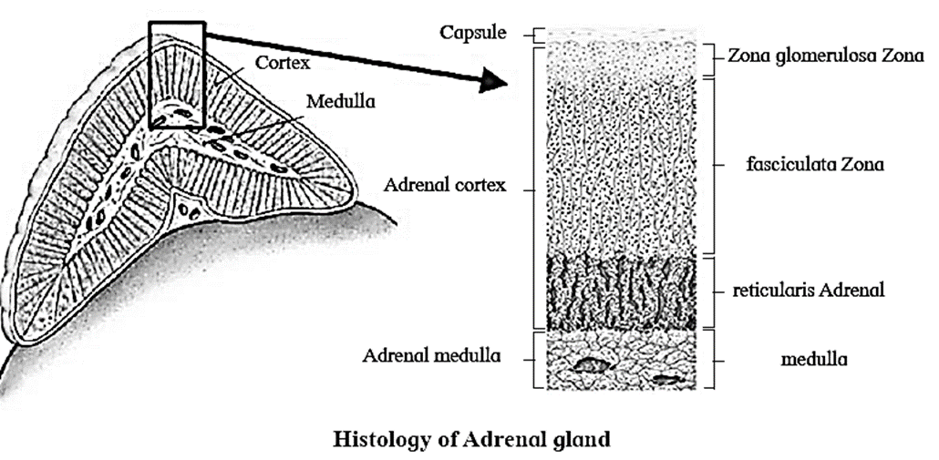
Three concentric regions of adrenal cortex :
Three concentric regions of adrenal cortex :
(i) Zona glomerulosa : Location : Outer thin cortical area
- It secretes Mineralocorticods.
- They are released for regulating sodium and potassium ion concentration. They regulate salt-water balance, blood volume and blood pressure.
- Aldosterone (salt retaining hormone) is the main mineralocorticoid. It balances Na-K levels.
(ii) Zona fasciculata : Location : Middle thick zone of cortex.
- It is responsible for secretion of Glucocorticoids like cortisol.
- It regulates metabolism of carbohydrates, proteins and lipids.
- Cortisol is an important glucocorticoid. It is responsible for increase in blood glucose level. It is also immuno suppressive. It suppresses synthesis of antibodies. So it is used in treatment of allergy.
- It prepares animals to face emergencies in nature.
(iii) Zona reticularis : Location : Inner thin zone of cortex.
- It is responsible for production of sex corticoids (Gonadocorticoids).
- In males, they have a role in development and maintenance of external sex characters.
- Excess sex corticoids in female causes adrenal virilism and hirsutism (excess hair on face) while in males it causes gynaecomastia i.e. enlarged breast. In males it causes gynaecomastia.
- Androgens and estradiols are the produced by the adrenal cortex.
[collapse]
Disorders related to Adrenal cortex :
Disorders related to Adrenal cortex :
- Hyposecretion : Hyposecretion of mineralocorticoids and glucocorticoids is responsible for Addison’s disease. Symptoms of this disease are low blood sugar, low Na+ and high K+ concentration in plasma, increased loss of Na+ and water in urine. It leads to weight loss, weakness, nausea, vomiting and diarrhoea.
- Hyper secretion : Hyper secretion of glucocorticoids produces Cushing’s disease. It leads to high blood sugar level, excretion of glucose in urine, rise Na+ in blood volume, high blood pressure, obesity and wasting of limb muscles.
[collapse]
Pancreas :
- Develops from endoderm.
- It is heterocrine i.e. both exocrine and endocrine gland.
- The exocrine part is pancreatic acini. The cells of acini secrete pancreatic juice containing digestive enzymes like trypsinogen, chymotrypsinogen, etc.
- Endocrine cells of pancreas form groups of cells called Islets of Langerhans.
There are four kinds of cells in islets of Langerhans which secrete hormones.
- Alpha (a) cells (20%) secrete glucagon. Glucagon is a hyperglycemic hormone. It stimulates liver for glucogenolysis and increases the blood glucose level.
- Beta (b) cells (70%) secrete insulin. Insulin is a hypoglycemic hormone. It stimulates liver and muscles for glycogenesis. This lowers blood glucose level.
- Delta (d) cell (5%) secrete somatostatin. Somatostatin inhibits the secretion of glucagon and insulin. It also decreases the gastric secretions, motility and absorption in digestive tract. In general it is a growth inhibiting factor.
- PP cells or F cells (5%) secrete pancreatic polypeptide (PP), which inhibits the release of pancreatic juice.
Disorder related to pancreas :
Disorder related to pancreas : Diabetes mellitus
- Hyperglycemia i.e. It leads to increased blood glucose level.
- Cause : Under activity of Beta cells, which results in reduced secretion of insulin.
Types of diabetes :
TYPE-1 diabetes : Insulin dependent diabetes mellitus (IDDM)
TYPE-2 diabetes : Non insulin dependent diabetes mellitus (NIDDM).
- Diabetes causes glucosuria, excessive urination and dehydration of body tissues, degradation of fats and increase in formation of ketone bodies (ketosis).
- Administration of insulin lowers blood glucose level.
[collapse]
Gonads :
Gonads are sex organs (the testes and the ovaries).
(1) Ovaries :
Ovaries : They produce harmone
(i) Estrogen :
- These are secreted by developing follicle.
- Estradiol is the main oestrogen.
- It is responsible for secondary sexual characters in female.
(ii) Progesterone :
- It is secreted by corpus luteum of the ovary after ovulation.
- This hormone is essential for thickening of uterine endometrium, thus preparing the uterus for implantation of fertilized ovum.
- It is responsible for development of mammary glands during pregnancy.
- It inhibits uterine contractions during pregnancy.
(iii) Relaxin :
- It is secreted by the corpus luteum of the ovary at the end of gestation period.
- It relaxes the cervix of the pregnant female and ligaments of pelvic girdle for easy birth of young one.
(iv) Inhibin :
- It is secreted by the corpus luteum.
- It inhibits the FSH and GnRH production.
[collapse]
(2) Testes :
Testes :
Testes secrete male sex hormones called androgens such as testosterone.
Testosterone :
- It is secreted from interstitial cells or Leydig cells by the influence of luteinizing hormone (LH).
- Rise in testosterone level in blood above normal inhibits LH secretion.
- It is also responsible for appearance of secondary sexual characters such as facial and pubic hair, deepening of voice, broadening of shoulders, male aggressiveness, etc.
[collapse]
Placenta :
- Temporary endocrine source in pregnant women which forms intimate connection between foetus and uterine wall of the mother for physiological exchange of the materials.
- During pregnancy, placenta secretes hormones such as estrogen, progesterone
- HCG (Human Chorionic Gonadotropin) and human placental progesterone.
Diffused endocrine glands :
Diffused endocrine glands :
Gastro-intestinal tract : Certain cells, of gastrointestinal mucosa are endocrine in function. Their hormones play vital role in digestive processes and flow of digestive juices.
- Gastrin : It stimulates gastric glands to produce gastric juice.
- Secretin : It is responsible for secretion of pancreatic juice and bile from presence and liver.
- Cholecystokinin CCK/ Pancreozymin PZ : This hormone stimulates the pancreas to release its enzymes and also stimulates gall bladder to release bile.
- Entero-gastrone / Gastric inhibitory peptide (GIP) : It slows gastric contractions and inhibits the secretion of gastric juice.
Kidney :
- Hormones of kidney - renin, erythropoietin and calcitriol (calcitriol is the active form of vitamin cholecalciferol -D3).
- Renin along with angiotensin helps in maintaining the blood pressure in the renal artery by vasoconstriction.
- Erythropoietin stimulates erythropoiesis.
- Calcitriol helps in absorbing calcium from the stomach.
Heart :
- The atrial wall of the heart secrete a peptide hormone known as atrial natriuretic factor (ANF).
- Increases sodium excretion [natriuresis] along with water by kidneys.
- Reduces blood pressure by lowering blood volume.
- When the blood pressure increases, ANF hormone is secreted.
- It causes dilation of the blood vessels.
- Blood then can easily flow with lesser resistance and hence BP decreases.
[collapse]
Hormone therapy/HT :
- Use of hormones in medical treatment.
- Required for the patients during pregnancy, menopause, osteoporosis, growth hormone deficiency, insulin resistance, cancer, etc.
<<-Previous Part
We reply to valid query.