|
Topics to be Learn : Part-1
- Introduction
- Immunity
- Structure of antibody
- Common human diseases
Topics to be Learn : Part-2
- Adolescence
- Addiction
- Drugs abuse
|
Introduction :
Health : According to the World Health Organization (WHO), health is defined as the state of complete physical, mental and social wellbeing and not merely the absence of disease or infirmity.
Health is metabolic and functional efficiency of living organisms.
Hygiene : Hygiene is a science of rules of health.
- To achieve good health, it is therefore, necessary that we have Hygienic balanced diet, clean drinking water, personal and community hygiene, regular physical exercise, knowledge about diseases and their effect on body, proper disposal of wastes and control of vectors.
Human Body Health System :
Human Body Health System :
- All human beings are exposed to various foreign bodies, including infectious agents like bacteria, viruses, etc. which are called pathogens.
- But human body can resist almost all types of these pathogens.
- For this purpose, there is immune system which protects us from various infectious agents.
- There is resistance and prevention of the damage or disease, through our defence mechanisms.
- Thus, despite constant exposure to variety of pathogens, most of us remain healthy.
[collapse]
Immune system : The ability to resist almost all types of these foreign bodies is possible due to immune system. It protects human beings from various pathogens or infectious agents.
Resistance : The ability to prevent the damage or disease, through our defence mechanism.
Immunity :
- Edward Jenner : Started the concept of immunity. Cowpox vaccine was developed by him.
- Immunity : Immunity is the resistance exhibited by the host towards infections caused by pathogens and their products.
- Immunology : Study of immune system, immune responses to foreign substances and their role in resisting infection by pathogens.
- Antigen : Any foreign substance invading body and capable of stimulating an immune response.
- Antibody : The protective chemicals produced by immune cells in response to antigens are called antibodies.
Types of immunity:
There are two basic types of immunity, viz. innate immunity and acquired immunity.
Innate immunity :
- Innate immunity is natural, inborn immunity, which helps the body to fight against the invasion of microorganisms.
- Innate immunity is non-specific because it does not depend on previous exposure to foreign substances.
- Innate immunity mechanisms consist of various types of barriers such as anatomical barriers, physiological barriers, phagocytic barriers and inflammatory barriers. They prevent entry of foreign agents into the body.
Inborn immunity which is non-specific and not affected by immunization.
Barriers of innate immunity :
The barriers of innate immunity are as follows :
(i) Epithelial surface : Different ways in which epithelial surface offers the innate immunity are :
- Skin and mucous covering, when intact protects the body against the invasion by pathogens. The healthy skin has bactericidal activity due to the salts present in drying sweat.
- Sebaceous glands in the skin produce secretions and long chain of fatty acids. These are bactericidal and fungicidal.
- Respiratory tract is provided with mucosa which prevents entry of microorganisms to a large extent.
- The inhaled particles are arrested through hair in the nasal passage.
- The particles that pass beyond nasal passage are caught by mucus lining the epithelium.
- They are swept back to pharynx. Then they are either swallowed or coughed out.
- The cough reflex is an important defence mechanism of respiratory tract.
- There is saliva in the mouth which has inhibitory effect on microorganisms. Gastric secretions has acidity and hence microorganisms are destroyed in stomach.
- The flushing action of urine eliminates bacteria from the urethra. Semen too has antibacterial substances, e.g. Spermine and zinc.
(ii) Antimicrobial substances in blood and tissues :
- The complement system with 30 serum proteins in blood in an inactive state.
- Complement cascade : Microbial pathogens activates the ‘Complement cascade’ to eliminate pathogens.
- The interferons —>cytokines (soluble proteins) released by cells infected with viruses.
- WBC to stimulate other cells.
(iii) Cellular factors in innate immunity :
- Phagocytic cells ingest and destroy the pathogens.
- This is natural defence against the invasion of pathogenic microorganisms and other foreign particles in blood and tissues.
- Phagocytic cells are of two types, viz. microphages and macrophages. They can remove foreign particles that enter the body.
- Natural killer (NK) cells is a class of lymphocytes which carry out important and non-specific defence against viral infections and tumours.
(iv) Fever :
- Natural defence mechanism by raising body temperature to accelerate the physiological processes.
- Interferon production stimulated by fever.
(v) Acute phase proteins (APPs) :
- Acute phase proteins are involved in innate immune mechanism.
- When there is an infection or injury, it leads to a sudden increase in concentration of certain plasma proteins, which are called acute phase proteins or APPs.
- These include C Reactive Protein (CRP), Mannose binding protein, Alpha-1-acid glycoprotein, Serum Amyloid P. etc.
- APPs enhance host resistance, prevent tissue injury and promote repair of inflammatory lesions.
[collapse]
Acquired immunity :
- The immunity that an individual acquires during his life is called acquired immunity or adaptive immunity or specific immunity.
- Specific or adaptive immunity acquired during lifetime due to infections
- It helps the body to adapt by fighting against specific antigens hence it is called adaptive immunity. Since it is produced specifically against an antigen, it is called specific immunity.
- Acquired immunity takes long time for its activation.
- This type of immunity is seen only in vertebrates.
- It involves formation of antibodies from the destruction of foreign antigens
- Unique features of acquired immunity are specificity, diversity discrimination between self and non-self and memory.
Types of acquired immunity :
Types of acquired immunity : Active immunity and Passive immunity
(i) Active immunity :
- Active immunity is produced in response to entry of pathogens and their antigenic stimuli.
- Active immunity is the long lasting immunity.
- In active immunity, the body produces its own antibodies.
- Natural acquired active immunity is obtained due to infections by pathogens.
- Artificial acquired active immunity is obtained through vaccinations. These vaccines contain dead or live but attenuated pathogens.
(ii) Passive immunity :
- Passive immunity is produced due to antibodies that are transferred to the body.
- Passive immunity is short-lived immunity.
- In passive immunity, antibodies are given to the body from outside.
- Natural acquired passive immunity is obtained through antibodies of mother transmitted to baby by placenta or colostrum.
- Artificial acquired passive immunity is also obtained through vaccinations, but here the vaccines contain the readymade antibodies which are prepared with the help of other animals such as horses.
[collapse]
Cells of Immune System :
Cells of Immune System:
There are two main types of cells involved in the working of Immune system. They are (a) Lymphocytes and (b) Antigen Presenting cells.
(a) Lymphocytes :
Lymphocytes are the main cells of the immune system. They, like the other blood corpuscles, arise from the stem cells, the haemocytoblasts, present in liver of the foetus and in the bone marrow in adult.
Bursa! / B-lymphocyte :
- Differentiation in the gut associated bursal lymphoid tissues, e.g. Tonsils, Peyer’s patches.
- Form humoral or antibody mediated immune system (AMIS).
Thymus / T-lymphocytes :
- Differentiation in the thymus.
- Form cell-mediated immune system (CMIS).
Mechanism of response of T-lymphocytes to antigens : On coming in contact with an antigen, a T-lymphocyte forms clones of T-cells which are similar but they perform different functions. The clone has four types of T-lymphocytes:
Four types of T-lymphocytes :
- Helper T-cells : produce lymphokines f.
- Killer T-cells or Cytotoxic T-cells : directly attack and destroy invading microbes, secrete perforins.
- Suppressor T-cells : Suppress entire immune system against attack on the own body cells.
- Memory T-cells : These previously sensitized cells that remember the antigen.
Mechanism of action of B-lymphocytes to antigens:
- B–lymphocytes are sensitized directly by both antigens as well as by helper T-cells.
- Activated B-lymphocyte multiplies very fast to produce clone of plasma cells and memory B-cells.
- The plasma cells produce specialized glycoproteins, called antibodies which are circulated through body fluids (humor) like blood and lymph.
- The antibody molecules may bind to a cell membrane or they remain free.
The free antibodies have three main functions:
- Agglutination of particulate matter, including bacteria and viruses. The immobilized mass is then engulfed by phagocytes.
- Opsonisation or coating of bacteria to facilitate their subsequent phagocytosis by macrophages.
- Neutralization of toxins released by bacteria e.g. tetanus toxin.
Each antibody is specific for a particular antigen.
(b) Antigen Presenting cells :
- Antigen presenting cells engulf invading pathogens and process the antigens.
- Deliver a stimulatory signal that is necessary for activation of helper T-cell.
[collapse]
Vaccination :
Vaccination : Administration of inactivated pathogen or antigenic protection of particular pathogen for protecting against a particular pathogen, is called vaccination.
Vaccine : Inactivated pathogen or antigens of a specific disease.
- Vaccines are prepared from inactivated pathogen, in the form of protein or sugar from pathogen or dead form of pathogen or toxoid from pathogens or attenuated pathogen.
- These when they are administered to a person to protect against a particular pathogen, it is called vaccination.
- Vaccination ’teaches’ the immune system to recognize and eliminate pathogenic organism. Because, already in the body the vaccine is injected and body has made antibodies in response to it. Thus, body is prepared before the attack, if at all it is exposed to pathogen.
- Thus, it is an important form of primary prevention, which reduces the chances of illness by protecting people. It works by exposing the pathogen in a safe form.
- Vaccinations control spread of diseases like measles, polio, tetanus and whooping cough that once threatened many lives.
- Vaccination controls the epidemic outbreak of diseases, if all the people are pre-vaccinated.
- Some hazardous diseases like small pox and polio have been completely eradicated by the vaccination.
Structure of Antibody :
Structure of Antibody:
Antibodies are glycoproteins which are highly specific to specific antigens.
- They are produced by plasma cells. Plasma cells are in turn formed by B-lymphocytes.
- About 2000 molecules of antibodies are formed per second by the plasma cells.
- Antibody is a ‘Y’-shaped molecule. It has four polypeptide chains, two heavy or
- H-chains and two light or L-chains.
Click here to View Figure-1
- Disulfide bonds (-s-s-) hold the polypeptide chains together to form a ‘Y’-shaped structure.
- The region holding arms and stem of antibody is termed as hinge. Each chain of the antibody has two distinct regions, the variable region and the constant region.
- Variable regions have a paratope which is an antigen-binding site. This part of antibody recognizes and binds to the specific antigen forming an antigen-antibody complex.
- Antibodies are called bivalent as they carry two antigen binding sites.
[collapse]
Formation of antigen-antibody complex :
Formation of antigen-antibody complex :
- Between antigen and antibody there is specificity.
- Each antibody is specific for a particular antigen.
- On the antigens there are combining sites which are called antigenic determinants or epitopes.
- Epitopes react with the corresponding antigen binding sites of antibodies which are called paratopes.
- The antigen binding sites are located on the variable regions of the antibody. Variable regions have small variations which make each antibody highly specific for a particular antigen.
- Owing to variable region the antibody can recognize the specific antigen.
- Antibody thus binds to specific antigen in a lock and key manner, forming an antigen-antibody complex.
Click here to View Figure-2
[collapse]
Antigen on Blood Cells : Antigens (A, B,D) on the surface of human red blood cells : responsible for different blood groups.
- There are about 30 known antigens on the surface of human red blood cells. They decide the type of blood group such as ABO, Rh, Duffy, Kidd, Lewis, R MNS, Bombay.
- The different blood groups are determined genetically due to presence of a particular antigen.
- Landsteiner found two antigens or agglutinogens on the surface of human red blood cells which are named as antigen A and antigen B.
- There is another antigen called Antigen D which decides the Rh status of the blood. If Antigen D is present, the person is said to be RH positive and when it is lacking, the person is Rh negative.
- These antigens are responsible for types of blood group and the specific transfusions.
- Antigens present on the RBCs and antibodies present in the serum can cause agglutination reactions if they are non-compatible.
- Therefore, at the time of transfusion blood groups are checked properly.
Blood group systems : ABO, Rh, Duffy, Kidd, Lewis, P, MNS, Bombay blood group, etc.
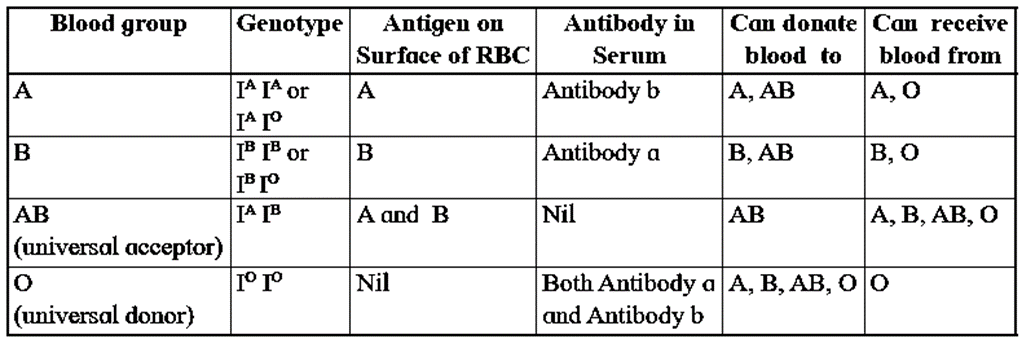
ABO blood group system :
ABO blood group system :
- In ABO system, the blood groups are determined by the antigen present on the surface of red blood cells.
- The blood groups are of four types, viz A, B, AB and O.
- In person with blood group A there is antigen A on the surface of their red blood cells (RBCs) and antibodies ‘b’ in their plasma.
- In person with blood group B there is antigen ‘B’ on the surface of their red blood cells (RBCs) and antibodies ‘a’ in their plasma.
- In person with blood group AB there are both antigens A’ and ‘B’ on the surface of their RBCs and no antibodies in their plasma.
- In person with blood group ‘O’ there are no antigens ‘A’ and ‘B’ on the surface of their RBCs but have both ‘a’ and ‘b’ antibodies in their plasma.
- During blood transfusion compatibility of blood has to be taken into consideration.
- Person with ‘O’ blood group is called universal donor while the person with ‘AB’ blood group is called universal recipient.
- Individuals with blood group O can donate blood to anyone, while those individuals with blood group AB can receive blood from any person.
ABO Blood groups in man chart :
Click here to View Chart
- Karl Landsteiner : Discovered A, B and O blood groups in 1900
- Decastello and Sturli discovered blood group AB in 1902.
[collapse]
Rh factor :
- Rh factor is the term adapted from Rhesus monkey.
- In rhesus monkey, there is antigen D on the surface of their RBCs.
- Landsteiner and Wiener discovered this antigen and termed it as Rh factor.
- Persons having Rh factor or D antigen are called Rh positive while those lacking D antigen or Rh factor are called Rh negative.
- Rh (D) antigen induces a strong immunogenic response when introduced into Rh -ve individuals.
- Haemolytic diseases of the newborn (HDN), or erythroblastosis foetalis occurs when an Rh—ve mother conceives Rh+ve foetus.
Common Human Diseases :
Disease : Disease is defined as condition of disturbed or deranged functioning of one or more organs or organ systems of the body, caused due to infections, defective diet or heredity.
All human diseases are classified into two types : Congenital diseases and Acquired diseases.
Congenital diseases :
- Congenital diseases are present from birth;
- Caused by genetic abnormality or metabolic disorder.
- They may be permanent and were practically incurable.
- Modern research has helped to cure some inborn diseases through gene therapy, enzyme replacement therapy, etc.
Acquired diseases :
Diseases which are developed after the birth of an individual are called acquired diseases.
These are of two types, viz. (a) Communicable or infectious diseases and (b) Non-communicable or Non-infectious diseases.
Communicable or infectious diseases :
Communicable or infectious diseases :
- Diseases transmitted from infected person to healthy person are called communicable or infectious diseases.
- Communicable diseases spread through pathogens.
- Communicable diseases are not inherited from parental generation to offspring.
- Vectors play the major role in spreading disease from one person to another.
- Treated by conventional methods using antibiotics and other drugs.
- Diseases are acute which develop suddenly due to infections.
- E.g. Pneumonia, Tuberculosis, AIDS, Typhoid, Cholera, Malaria.
[collapse]
Non-communicable or Non-infectious diseases :
Non-communicable or Non-infectious diseases :
- Diseases that are not passed from one person to other are non-communicable or non-infectious diseases.
- Non-communicable diseases do not spread through pathogens. ’
- Non-communicable diseases like cancer can be from parental generation to offspring.
- Caused due to allergy, illness, malnutrition or abnormalities in cell proliferation, changes in lifestyle, environment play a significant role.
- Treated conservatively for a long time or surgically.
- Diseases are chronic which develop and persist for a long time.
- E.g. Cancer, Rickets, Allergies, Kwashiorkor, Diabetes, Heart disease, etc.
[collapse]
Examples of Diseases :
(1) Malaria (Type-Protozoan ) :
Mode of transmission :
- Malaria parasite is transmitted through the female Anopheles mosquito and hence it is known as mosquito-borne disease. Mosquito acts as a vector.
Causative organisms :
- Disease caused by protist - Plasmodium. There are four species of Plasmodium, viz. P. vivax, P. falciparum, P. ovale and P. malariae which transmit malaria.
Symptoms :
Symptoms :
- Fever accompanied by shivering, Joint pain or arthralgia, Vomiting, Anaemia caused due to rupture of RBCs or haemolysis, Haemoglobinuria, Retinal damage, Convulsions etc.
- Cyclical occurrence of sudden coldness followed by rigor and then fever and sweating lasting for four to six hours. This is called a classic symptom of malaria.
- Splenomegaly or enlarged spleen, severe headache, cerebral ischemia, hepatomegaly, i.e. enlarged liver, hypoglycaemia and haemoglobinuria with renal failure may occur in severe infections.
[collapse]
Diagnosis and Treatment :
- Blood smear testing.
- Nucleic acid amplification technique.
- ACT therapy. (5 different ACTs).
- Includes various combinations of artesunate, sulfadoxine, pyrimethamine, etc. and quinine.
Preventive measures :
- Transmission of malarial parasite can be reduced by preventing mosquito bites.
- Therefore, mosquitoes should be controlled or totally eradicated.
- This can be done by using of mosquito nets and insect repellents.
- Mosquito control measures such as spraying insecticides inside houses and draining stagnant water where mosquitoes lay their eggs.
- The mosquito larvae can be eradicated by releasing Gambusia fish which can feed upon these larvae.
- Vaccine against malaria is also under preparation.
(2) Amoebiasis or Amoebic dysentery (Type-Protozoan)
Mode of transmission :
Amoebiasis is usually transmitted by the following ways :
- The faecal-oral route.
- Through contact with dirty hands or objects.
- By anal-oral contact.
- Through contaminated food and water.
Causative organisms :
- Amoebiosis is spread through ingestion of the cyst form of Entamoeba histolytica. This is a commensal organism.
- Cyst is a semi-dormant and hardy structure found in faces of infected person.
- Non-encysted amoebae are called trophozoites. The trophozoites die quickly after leaving the body but may also be present in faces.
- Trophozoites are rarely the source of new infections.
- The infection may remain asymptomatic for many days as Amoeba can remain latent in the gastrointestinal tract. .
Symptoms :
Symptoms :
- Amoebiasis shows following common symptoms :
- Diarrhoea, flatulence, stool with mucus and abdominal pains (cramps) are common.
- Stool sticky with mucus and blood.
- Amoebae form cysts in the liver, in such case there is hepatomegaly, i.e. enlargement of liver.
- Liver shows amoebic liver abscess accompanied with fever and pain in right side of the abdomen.
[collapse]
Diagnosis and Treatment :
- Microscopic examination of the stool
- Metxonidazole and Timidazole.
Preventive measures :
Prevention of amoebiasis is to be done at two levels, viz. at home and at endemic level.
Prevention of the spread of amoebiasis at the home level :
- Washing hands with soap and water after using the toilet or changing a baby’s diaper and before handling and eating food.
- Cleaning bathrooms and toilets properly with germicides.
- Avoiding raw vegetables when in endemic areas where they are grown in soil fertilized by human faeces.
- Boiling and purifying the drinking water.
Prevention of the spread of amoebiasis at endemic level :
- Avoiding consumption of street foods especially in public places.
- Following good sanitary practice, as well as using proper sewage disposal or treatment.
- E. histolytica cysts are usually resistant to chlorination; therefore sedimentation and filtration of water supplies are necessary to reduce the incidence of infection.
- Avoiding shared towels or face washers.
(3) Ascariasis (Type-Helminth) :
Mode of transmission :
- Contaminated food and drinks which has eggs of Ascaris.
- Hatching of eggs occurs in intestine of host.
- Larvae enter various organs through circulation. As adult they settle in digestive system of host.
Causative organisms :
- Ascaris lumbricoides
- Endoparasite, roundworm or nematode
Symptoms :
Symptoms :
- After infection by Ascaris lumbricoides there is appearance of eggs in stools in 60 — 70 days.
- In larval ascariasis, symptoms are seen in 4 - 16 days after infection.
- The final symptoms are gastrointestinal discomfort, colic and vomiting, fever and appearance of live worms in faeces.
- Some patients may have pulmonary symptoms. Inflammation of alveolar walls is seen. This is known as pneumonitis.
- Some may show neurological disorders during migration of the larvae.
- Loss of appetite which reflects in weight loss.
- A bolus of worms may obstruct the intestine.
- Larvae that migrate may also cause eosinophilia, i.e. increase in number of eosinophils.
[collapse]
Diagnosis and Treatment :
- Anti-helrninthic drugs like Piperazine. Mebendazole, Lcvamisole, Pyrantel
- Microscopic examination of the stool.
Preventive measures :
Prevention of ascariasis can be done by adopting the following measures :
- Use of proper toilet facilities.
- Safe disposal of excreta.
- Protection of food from dirt and soil.
- Washing of vegetables before cooking and avoiding eating raw, unwashed vegetables and fruits.
- Hand washing and use of safe food.
- Observing personal hygiene.
- Use of pharmaceutical drugs such as Mebendazole and Albendazole can kill Ascaris.
(4) Filariasis or Elephantiasis (Type-Helminth Nematode endo parasite)
Mode of transmission :
Mode of infection, i.e. transmission :
- The parasite Wuchereria bancrofti is transmitted from a patient to other normal human being by female Culex mosquito.
- The filarial larvae leave mosquito body and arrive on the human skin where they penetrate the skin and enter inside.
- They undergo two moultings to become adults. Later they settle in the lymphatic system. They incubate for about 8- 16 months.
- When they settle in lymphatic system, thin infection is called lymphatic filariasis.
- The worms start infecting lymphatic circulation resulting into enlargement of lymph vessels and lymph nodes. The extremities like legs or limbs become swollen which resembles elephant legs.
- Therefore it is called elephantiasis.
- This condition is lymphoedema, i.e. accumulation of lymph fluid in tissue causing swelling.
Causative organisms :
- Lymphatic Filariasis- Wuchereria bancrofti, Brugia malayi, Brugia timori.
- Subcutaneous Filariasis- Loa loa. Mansonella spp.
- Serous cavity Filariasis -Mansonella spp.
Symptoms :
Symptoms :
- As the lymphatic drainage does not take place, there is oedema with thickening of skin and underlying ’tissue.
- Extremities like legs. arms, breasts, scrotum, etc. are affected by nematode causing lymphatic filariasis, i. e. Wuchereria bancrofti.
- Lymph vessels and lymph nodes are enlarged and swollen.
- Elephantiasis is seen in which limbs are swollen like legs of elephant.
- Lymphoedema, i.e. accumulation of lymph fluid is seen in tissue causing swelling.
- Hydrocele condition develops in which testis are enlarged due to accumulation of lymphatic fluid in testis.
[collapse]
Diagnosis and Treatment :
- For the patient, diethyl-carbamazine citrate is the drug used for twice a day for three weeks. Thereafter for five days every six months the same treatment is repeated. This becomes effective against filarial worms.
Preventive measures :
- Mosquito eradication should be done for controlling filariasis.
- In the areas with mosquitoes, avoid mosquito bite by using mosquito nets and insect repellents.
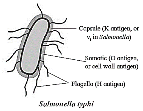
(5) Typhoid (Type-Bacterial) :
(5) Typhoid (Type-Bacterial) :
Mode of transmission :
- Typhoid is an infective disease caused by Gram—ve bacterium, Salmonella typhi.
- Food and water borne disease.
- Housefly and cockroaches can also transfer the bacteria from faeces to food.
- Poor hygiene habits.
- Poor sanitation.
Causative organisms :
- Salmonella typhi. (Gram negative bacteria)
- Intestinal lumen of infected person lodges it.
- The bacterium has “O” — antigen, which is a lipopolysaccharide (LPS), present on surface coat and its flagella has — antigen. Thus it becomes pathogenic.
Click here to View Figure-4
Symptoms :
- Prolonged high fever (104°F)
- General nausea, fatigue, head ache.
- Abdominal pain. Constipation or diarrhoea.
- Rose-coloured rash on skin.
- White coat on tongue. cough.
- Anorexia (loss of apetite).
- Breathlessness. Irregular heartbeats, haemorrhage, if neglected .
Diagnosis and Treatment :
- Widal test: diagnosis of typhoid.
- Treatment of typhoid involves surgical removal of gall bladder in severe cases.
- Chlorornycetin -antibiotics
Preventive measures :
- Cleanliness. Clean and safe food.
- Avoiding roadside food.
- Two available vaccines are : (1) Oral: Ty21a vaccine, (2) Injectable: typhim vi and typherix
[collapse]
6) Pneumonia (Type- Bacterial, Viral, Physical reasons, Burns) :
(6) Pneumonia (Type- Bacterial, Viral, Physical reasons, Burns)
Mode of transmission :
- Direct contact from person to person
- Droplets infection.
- Shared clothes and utensils
Causative organisms :
- Virus species: Influenza virus. adenovirus. para influenza, Respiratory Syncytial Virus (RSV).
- Bacteria: Streptococcus pneumonia
- Fungal species: ‘Pneumocystis jirovecii, Pneumocystis carinii.
Symptoms :
- Cough, Yellow or greenish sputum or phlegm.
- High fever.
- Shortness of breath (Dyspnea).
- Deep breath or coughing along with chest pain.
- Loss of appetite, fatigue, headaches, Vomiting, joint pains and muscle aches.
- Inflammation of respiratory passage and lungs
Diagnosis and Treatment :
- Treatment as per pathogen type.
- Bacterial pneumonia: antibiotics -Benzyl penicillin. Ampicillin and Chloramphenicol.
Preventive measures :
- Vaccination is important prevention in both children ancl adults.
- Vaccines against Haemophilus influenzae and Streptococcus pneurnoniae are given in first year of life.
[collapse]
(7) Common cold (Type-Viral) :
(7) Common cold (Type-Viral) :
Mode of transmission :
- Transmitted through droplet infections:
- Nasopharyngitis
- Acute viral rhinopharyngits
- Acute coryza or a cold
Causative organisms :
- Rhinoviruses and Corona-viruses
Symptoms :
- Cough. sore throat, runny nose and fever.
- Sneezing with nasal congestion, Conjunctivitis (red eyes)
- Muscle rashes, fatigue, headache
- Shivering and loss of appetite
Diagnosis and Treatment :
- No medicines.
- Painkiilers and paracetamol may help
- Homemade remedies more common.
Preventive measures :
- To keep away from persons having common cold
- Hand-wash using soap and water.
- Handkerchief to cover the nose and mouth during coughing and sneezing.
- Alcohol based hand sanitizer can also be used.
[collapse]
(8) Ring worm Dermatophytosis (Type-Fungal) :
(8) Ring worm Dermatophytosis (Type-Fungal) :
Mode of transmission :
- Sharing of clothes, comb of infected person.
- Close contact with infected person
Causative organisms :
- Fungal species: Trichophyton and Microsporum.
Symptoms :
- Enlarged, red rings on skin.
- Intense itching.
- Appearance of dry, scaly lesions on various parts of the body
- Infection to nails turning them thick, discoloured and disfigured.
- The fungal infection that usually begins between the toes.
Diagnosis and Treatment :
- Physical examination
- Drugs: Nystatin. fluconazole, itraconazole, etc.
Preventive measures :
- No close contact
- No sharing of clothes and Sport equipment.
- Washing clothes in hot water with fungicidal soap
[collapse]
(9) Dengue (Type-Viral) :
(9) Dengue (Type-Viral) :
- Dengue is a viral disease causing high fever.
- It is a painful, debilitating vector-borne disease.
Mode of transmission :
- Vector of Dengue virus is female Aedes mosquito. The mosquito takes up the dengue virus when it sucks blood of a person suffering from dengue.
- The spread of dengue is not directly from one person to another person.
Causative organisms :
- Four closely related dengue viruses.
Symptoms :
- High fever
- Vomiting
- Severe headache
- Decrease in Platelet count
Diagnosis and Treatment :
- Blood test.
- Physical examination
Preventive measures :
- Mosquito control.
- Water should not remain stagnant in manmade containers.
[collapse]
(10) Cancer:
Abnormal, uncontrolled and purposeless division of cells may lead to the formation/ development of mass of undifferentiated cells i.e. tumor. When tumor is malignant, it is described as cancer and has ability to invade other tissues.
- Neoplasm : Masses of tissue which form lumps due to uncontrolled cell division.
- Oncologists : Oncologists are the physicians and researchers who specialize in the study, diagnosis treatment and prevention of cancer.
Tumors may develop anywhere in the body. But all tumors are not cancerous.
Benign tumour and malignant tumour :
There are two types of tumors: benign or nonmalignant and cancerous or malignant.
Benign tumour :
- Benign tumour is localized and it does not spread to neighbouring areas.
- It is enclosed in connective tissue sheath.
- It compresses the surrounding normal tissue.
- It can be removed surgically.
- Except for brain tumour, benign tumours are usually not fatal.
- It do not show metastasis.
- Benign tumours are well differentiated.
- Benign tumours show slow and progressive growth.
Malignant tumour :
- Malignant tumour starts as local but spreads rapidly to neighbouring areas.
- It is not enclosed in connective tissue sheath.
- It invades and destroys the surrounding tissue.
- It need further treatment after removal.
- Malignant tumours are fatal.
- Malignant tumours show metastasis.
- Malignant tumours are poorly differentiated.
- Malignant tumours show rapid and erratic growth.
Click here to View Figure-5
[collapse]
Symptoms of cancer :
- Presence of lump or tumour.
- White patches in the mouth.
- Change in a wart or mole on the skin.
- Swollen or enlarged lymph nodes.
- Vertigo, headaches or seizures if cancer affect the brain.
- Coughing and shortness of breath if lungs are affected due to cancer.
Types of Cancer :
Types of Cancer : According to the tissue affected, the cancers are classified into five main types. These are as follows :
- Carcinoma : Cancer of epithelial tissue covering or lining the body organs is known as carcinoma. E.g. breast cancer, lung cancer, cancer of stomach, skin cancer, etc.
- Sarcoma: Cancer of connective tissue is called sarcoma. Following are the types of sarcoma : osteosarcoma (bone cancer), myosarcoma (muscle cancer), chondrosarcoma (cancer of cartilage) and liposarcoma (cancer of adipose tissue).
- Lymphoma: Cancer of lymphatic tissue is called lymphoma. Lymphatic nodes, spleen and tissues of immune system are affected due to lymphoma.
- Leukaemia : Leukaemia is blood cancer. In this condition, excessive formation of leucocytes take place in the bone marrow. There are millions of abnormal immature leucocytes which cannot fight infections. Monocytic leukaemia, lymphoblastic leukaemia, etc. are the types of leukaemia.
- Adenocarcinoma : Cancer of glandular tissues such as thyroid, pituitary, adrenal, etc. is called adenocarcinoma.
[collapse]
Causes of Cancer :
Causes of Cancer: Following carcinogenic factors are responsible for causing cancer.
- Chemicals: Many induce development of cancer. E.g. nicotine, caffeine, polycyclic hydrocarbons and products of combustion of coal and oil. Sex hormone and steroids, if given or secreted in excess, can cause cancer. E.g. Breast cancer.
- Radiation : Radiations such as X-rays, gamma-rays, cosmic rays, ultra-violet rays are carcinogenic.
- Viruses : Virus possessing oncogenes (v-one genes) are carcinogenic. E.g. EBV (Epestin burr virus), HPV (Human papiloma virus) are oncogenic viruses.
- Oncogenes : Cellular oncogenes (e-one genes) or proto-oncogenes can cause cancer. They are present in normal cells but if activated they lead to oncogenic transformation of cells.
- Addiction : Addictive substances like cigarette smoke, tobacco lead to cancer of mouth, lips and lungs. Alcohol can cause cancer of oesophagus, stomach, intestine and liver. Drugs like marijuana or anaerobic steroids can also cause cancer.
[collapse]
Cancer treatment :
Cancer treatment : Cancer treatment consists of combination of a number of therapies which are follows :
- Chemotherapy : Chemotherapy means giving certain anticancer drugs. These drugs check cell division by inhibiting DNA synthesis. But these are more toxic to cancerous cell than to normal cells. Chemotherapy shows side effects such as hair loss or anaemia.
- Radiotherapy : In addition to chemotherapy, radiations are given. The cancer cells are bombarded with the radiations from radioactive materials such as cobalt, iridium and iodine. The X-rays, gamma rays and charge particles are used to destroy the cancerous tissue or cells. They cause minimum damage to the surrounding normal tissue or cells.
- Surgery: Entire cancerous tissue or cells are removed surgically. E.g. breast tumour or uterine tumour. After removing the cancerous tissue, additionally other treatments are also given.
- Immunotherapy : For tackling with tumour, patients are given biological response modifiers such as a-interferon which activates their immune system to destroy the tumour.
- Supportive therapy : With supportive therapy, patient’s quality of life is increased. To treat symptoms of cancer and side effect‘; of cancer treatments, this therapy is used. This therapy varies depending upon condition of individual patient.
[collapse]
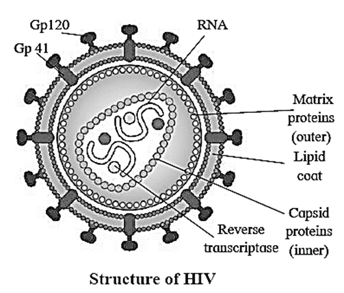
(11) AIDS :
AIDS : Acquired immuno deficiency syndrome, fatal and incurable illness caused by a retrovirus (ss RNA) called HIV (Human immunodeficiency virus).
- Body’s immune system is weakened increasing vulnerability causing many life threatening opportunistic infections, neurological disorders and malignancies.
Structure of HIV :
Structure of HIV :
Click here to View Figure-6
- Spherical, 100 to 140 nm in diameter, with centrally located two ss RNA molecules and reverse transcriptase enzymes.
- Covering of two layers of proteins.
- The outer layer is of matrix protein (p17) with additional layer of lipid.
- Impregnated with glycoprotein GP 12O and GP 41.
- Inner layer is capsid protein (p24)
- Replication of HIV in dividing
- T4 lymphocytes. They remain in latent state in lymphoid cells. “
[collapse]
Blood, semen and cerebrospinal fluid (CSF) show maximum concentration of HIV in infected person. Lesser extent seen in tears, milk, urine, saliva, cervical and vaginal secretions.
Transmission of virus :
- Unsafe sexual contact : Oral, vaginal and anal sex.
- Blood : Blood transfusions or sharing syringes, needles, etc.
- Transplacental (From mother to child during pregnancy via placenta) and by nursing mother through breast milk.
- Accidental needle injury, artificial insemination with infected semen and transplantation with infected organs.
- Through urine, tears, saliva, breast milk and vaginal secretions if these secretions enter passes in the body through injury.
Clinical manifestation of AIDS :
Clinical manifestation of AIDS :
There are four stages of clinical manifestations or symptoms of AIDS.
- Stage I : This is initial infection with the virus and formation of antibodies, usually 2-8 weeks after initial infection.
- Stage II : In this stage the person is asymptomatic carrier. Incubation takes place with a period ranging for 6 months to 10 years.
- Stage III : This is called AIDS related complex (ARC). In this stage, one or more of the following clinical signs are seen. E.g. Recurrent fever for longer than one month, fatigue, unexplained diarrhoea, night sweats, shortness of breath, loss of more than 10 per cent body weight, etc.
- Stage IV: This is the end stage in which patient shows full blown AIDS. Thus it is called the end stage of HIV infection. Life threatening opportunistic infections (like pneumonia, tuberculosis, Kaposi sarcoma, etc.) are easily caught during this period.
[collapse]
Diagnosis and Treatment :
Laboratory diagnosis :
- There are two tests for diagnosis of AIDS.
- First test is ELISA (Enzyme-Linked Immunosorbent Assay) which is used to detect the HIV antibodies.
- The second confirmatory test is Western Blot, which is used to weed out any false positive results. It is a highly specific test.
- It is based on detecting specific antibody to viral core protein and envelope glycoprotein.
Treatment of AIDS :
- AIDS cannot be cured.
- Antiretroviral drugs are used to reduce the viral load and prolong the life of HIV patient.
- E.g. Antiretroviral therapy (ART) uses drugs such as TDF (tenofovir), EFV (Efavirenz), Lamivudine (BTC), etc.
Preventive measures :
Preventive measures :
AIDS has no cure hence prevention is the best choice. The following steps help in preventing this dreadful disease.
- High risk group people should be educated about HIV transmission. They should never donate blood.
- Use of disposable needles and syringes should be done with proper disposal.
- Risky sexual habits should be avoided.
- Tooth brushes, razors, other articles that can become contaminated with blood should not be shared.
- Blood should be screened before receiving it.
- Routine screening of blood and semen donors, organ donors (kidney, liver, lung, cornea), and patients undergoing haemodialysis must be done.
- Pregnant women or those women who are contemplating pregnancy should be regularly screened.
[collapse]
Next Part- >>


We reply to valid query.